
New technology has been developed for obtaining same-time AFM and SEM imaging. With the combination of techniques, it is possible to obtain quick 3D topography, mechanical, electrical, and magnetic properties from AFM combined with 2D fast imaging from SEM at the same time [1].
Further, in microscopes equipped with energy-dispersive x-ray spectroscopy (EDS) detectors, it is possible to obtain specific surface chemistry information [1]. Focused ion beams (FIB) can be employed to dissect precise areas of samples. Finally, using the software processing capabilities both images are merged offering robust results in techniques that traditionally required different machines. Further applications are discussed in detail in a webinar series conducted by the creators on the reference list [2].
References
[1] LiteScope. Available at: <https://www.nenovision.com/litescopetm/why-afm-in-sem/> Access 05 Apr. 2022.
[2] AFM-in-SEM: future of complex and in situ correlative analysis. Available at: <https://www.youtube.com/watch?v=it2pyTmtO-A> 05 Apr. 22.
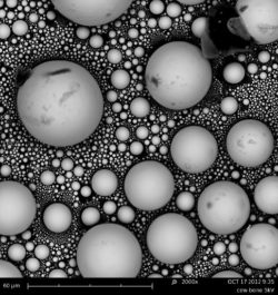
Header Image: SEM Images of cow bone CC BY-NC-ND 2.0.

This article was written by André Plath as part of an ongoing series of scientific communications written and curated by BioTrib’s Early Stage Researchers.
André is researching Boundary Lubrication of Fibrous Scaffolds at ETH Zürich, Switzerland.