What should be accepted in biomedical practice and research? New and old ethical dilemmas

Should young blood transfusions be allowed in rejuvenation processes? Is it ethical to pay for these transfusions? Should donors be remunerated? Is it ethical to increase the human life span by getting blood from younger donors transfused to older people? These questions stir up strong emotions in most of us. They represent not only ethical dilemmas but are within the boundaries of our scientific and philosophic understanding.

The history of human blood transfusions is odd. From early attempts with animals and bleeding to cure a series of ailments to our understanding of blood types, blood has stirred a lot of controversy. Bill Schutt brilliantly discusses some of these interesting stories in the lecture: What goes in: The strange story of blood transfusions available on Youtube.

With the new research on young blood transfusions to young patients (parabiosis), several effects were observed such as the reversion of aging-related degenerative diseases (1) and the increase of mice life span (2). This motivated the development of startups that quickly received huge sums of private funding (1). Since then, regulatory agencies such as the FDA have issued notes about the topic (3). In their note from 2019, FDA discusses: “Today, we’re alerting consumers and health care providers that treatments using plasma from young donors have not gone through the rigorous testing that the FDA normally requires in order to confirm the therapeutic benefit of a product and to ensure its safety. As a result, the reported uses of these products should not be assumed to be safe or effective.“ However, researchers are careful about these findings and recommend caution. The factors that play in these transfusions in animal studies are not clearly understood by the scientific community (3). Therefore, they cannot be translated into the complexity of humans. Given our little understanding, the regulation of research and the market of parabiosis is paramount. We should think together as a society on answers to the questions asked opening this short communication.

Read more:

(1)CORBYN, Zoe. Could young blood stop us from getting old? The Guardian: 2 Feb. 2020. Available at: https://www.theguardian.com/society/2020/feb/02/could-young-blood-stop-us-getting-old-transfusions-experiments-mice-plasma

(2)ZIMMER, Carl. Blood of young mice extends the life in the old? The New York Times, Jul. 2023. Available at: https://www.nytimes.com/2023/07/27/health/mice-blood-aging.html#:~:text=In%20the%20early%202000s%2C%20parabiosis,showed%20signs%20of%20accelerated%20aging

(3) GOTTLIEB, Scott. Statement from FDA Commissioner Scott Gottlieb, M.D., and Director of FDA’s Center for Biologics Evaluation and Research Peter Marks, M.D., Ph.D., cautioning consumers against receiving young donor plasma infusions that are promoted as unproven treatment for varying conditions. Available at: https://www.fda.gov/news-events/press-announcements/statement-fda-commissioner-scott-gottlieb-md-and-director-fdas-center-biologics-evaluation-and-0

SCHUTT, Bill. What goes in the strange story of blood transfusions? Available at: https://www.youtube.com/watch?v=DKs7iIwsU1c&t=1807s


This article was written by André Plath and Giulio Cavaliere as part of a series articles curated by BioTrib’s Early Stage Researchers.

André is one of BioTrib’s Early Stage Researcher‘s who is investigating Boundary Lubrication of Fibrous Scaffolds at ETH Zürich, Switzerland.

What is matrix-assisted chondrocyte implantation (MACI)?

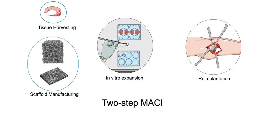
Header Image: Two-step Matrix Assisted Chondrocyte Implantation

Osteoarthritis is a condition associated with discomfort and significant challenges for the affected individuals. Pain management often involves the utilization of pain-relieving medications that can potentially lead to dependency [1]. Joint function restoration is achieved through the implementation of metal implants, although their durability is limited. Consequently, a secondary surgical procedure might be necessary as the patient ages. The use of these implants also presents additional complications such as inflammation induced by the gradual wear of microscopic components [2].

In response to these issues, chondrocyte implantation was introduced during the 1980s in Sweden. This medical procedure involves the extraction of cartilage or stem cells from the patient, which are then propagated within a supportive scaffold material (matrix) [3]. Subsequently, these multiplied cells are reintroduced into the patient’s body (Figure), contributing to tissue regeneration (two steps). An alternative method involves microfracture and scaffold implantation in one step, allowing cells to populate the material and facilitate cartilage reconstruction [3]. These newer techniques exhibit improved rates of success when compared to external cartilage growth [4].

However, a key challenge emerges when cartilage cells transform, losing their original properties. As a result, the newly generated tissue tends to become less lubricant. This phenomenon arises due to the limitations of the scaffold in replicating the intricate conditions found within living tissue [5]. The implications of this could involve detachment or accelerated wear of the implant, necessitating further surgical intervention, and potentially even the use of metal implants. Current materials utilized include foam structures derived from pork collagen or laser-etched plastics [6,7]. Nonetheless, promising advancements are being explored in the experimental phase, involving bio-based materials produced through techniques like 3D printing and fiber integration. One notable application of these innovations is the potential to postpone the need for a prosthesis, thereby offering a beneficial outcome, particularly for younger patients.


References

[1]          L.M. Billesberger, K.M. Fisher, Y.J. Qadri, R.L. Boortz-Marx, Procedural Treatments for Knee Osteoarthritis: A Review of Current Injectable Therapies, Pain Res. Manag. 2020 (2020) 1–11. https://doi.org/10.1155/2020/3873098.

[2]          O. Hussain, B. Ahmad, S. Saleem, Biomaterials for Artificial Knee Joint Replacement: A Review, Int. J. Mater. Eng. Innov. 14 (2023) 1. https://doi.org/10.1504/IJMATEI.2023.10048812.

[3]          G. Filardo, E. Kon, A. Roffi, A. Di Martino, M. Marcacci, Scaffold-Based Repair for Cartilage Healing: A Systematic Review and Technical Note, Arthrosc. J. Arthrosc. Relat. Surg. 29 (2013) 174–186. https://doi.org/10.1016/j.arthro.2012.05.891.

[4]          M.R. Steinwachs, J. Gille, M. Volz, S. Anders, R. Jakob, L. De Girolamo, P. Volpi, A. Schiavone-Panni, S. Scheffler, E. Reiss, U. Wittmann, Systematic Review and Meta-Analysis of the Clinical Evidence on the Use of Autologous Matrix-Induced Chondrogenesis in the Knee, CARTILAGE. 13 (2021) 42S-56S. https://doi.org/10.1177/1947603519870846.

[5]          A.R. Armiento, M.J. Stoddart, M. Alini, D. Eglin, Biomaterials for articular cartilage tissue engineering: Learning from biology, Acta Biomater. 65 (2018) 1–20. https://doi.org/10.1016/j.actbio.2017.11.021.

[6]          J.L. Carey, A.E. Remmers, D.C. Flanigan, Use of MACI (Autologous Cultured Chondrocytes on Porcine Collagen Membrane) in the United States: Preliminary Experience, Orthop. J. Sports Med. 8 (2020) 232596712094181. https://doi.org/10.1177/2325967120941816.

[7]          V.M. Mehta, S. Mehta, S. Santoro, R. Shriver, C. Mandala, C. Weess, Short term clinical outcomes of a Prochondrix® thin laser-etched osteochondral allograft for the treatment of articular cartilage defects in the knee, J. Orthop. Surg. 30 (2022) 102255362211417. https://doi.org/10.1177/10225536221141781.


This article was written by André Plath and Giulio Cavaliere as part of a series articles curated by BioTrib’s Early Stage Researchers.

André is one of BioTrib’s Early Stage Researcher‘s who is investigating Boundary Lubrication of Fibrous Scaffolds at ETH Zürich, Switzerland.

The Evolution of Total Knee Replacement: Tracing the Path of Surgical Advancement

Total Knee Replacement (TKR), also known as total knee arthroplasty (TKA), stands as one of the most remarkable achievements in modern medicine, revolutionizing the lives of countless individuals suffering from debilitating knee conditions. The history of total knee replacement is a journey through decades of innovation, surgical mastery, and persistent dedication to enhancing patients’ quality of life.

Early Endeavors and Conceptualization (19th to Early 20th Century)

The inception of total knee replacement can be traced back to the late 19th century when surgeons began to experiment with joint replacement procedures. However, it wasn’t until the early 20th century that the concept of total knee replacement started to take shape. One of the earliest documented efforts was by Themistocles Gluck in 1891, who proposed the idea of using ivory to replace a knee joint. This concept laid the foundation for future developments in knee replacement surgery.

Hinged Implants and Early Innovations (1950s – 1970s)

The mid-20th century witnessed significant advancements in orthopedic surgery, encouraging the development of hinged knee implants. In the 1950s, Dr. Leslie Gordon Percival Shiers successfully implanted a hinged knee joint, effectively treating severe arthritis. This marked a turning point, as surgeons began to explore alternatives to total knee fusion.

In the 1970s, the era of modern total knee replacement truly began. Dr. John Insall, along with his colleagues, developed the Insall-Burstein knee prosthesis, which aimed to reproduce natural knee kinematics. This design laid the groundwork for future innovations in implant design, implant fixation techniques, and surgical approaches.

Advancements in Implant Design and Surgical Techniques (1980s – 1990s)

The 1980s and 1990s brought remarkable progress in total knee replacement. Implant materials evolved, with surgeons experimenting with various combinations of metals and plastics to enhance durability and minimize wear. This period also saw the rise of minimally invasive surgical techniques, which aimed to reduce trauma, blood loss, and recovery time for patients.

Computer-Assisted Surgery and Personalized Implants (2000s – Present)

The turn of the millennium marked a shift toward computer-assisted surgeries and personalized implants. Computer navigation systems were introduced, enabling surgeons to achieve higher levels of accuracy in implant placement. Patient-specific implants, utilizing advanced imaging and 3D printing technologies, gained prominence, allowing for a more tailored approach to each patient’s anatomy.

Current Trends and Future Prospects

Total knee replacement continues to evolve. Researchers are exploring innovative materials, such as bioactive coatings and advanced polymers, to enhance implant longevity. Robotic-assisted surgeries have emerged, offering even greater precision and potentially improved outcomes.

Furthermore, the focus has extended beyond the surgical procedure itself to post-operative rehabilitation and patient education. Multidisciplinary approaches now involve physical therapists, pain management specialists, and psychologists, ensuring patients recover not only physically but also mentally.

In conclusion, the history of total knee replacement mirrors the remarkable progress of medical science and technology. From early conceptualizations to today’s state-of-the-art procedures, the journey has been marked by innovative ideas, persistent experimentation, and a commitment to enhancing patients’ lives. As we move forward, it’s likely that total knee replacement will continue to push the boundaries of what’s possible, providing renewed hope and mobility to individuals around the world.


References

  • Ranawat, History of total knee replacement. J South Orthop Assoc. 2002 Winter;11(4):218-26. PMID: 12597066.
  • Gunston, FH. Polycentric knee arthroplasty. Prosthetic simulation of normal knee movement: interim report.Clin Orthop Relat Res, 1973(94): p. 128-35.


This article was written by Elisa Bissacco as part of an ongoing series of scientific communications written and curated by BioTrib’s Early Stage Researchers.

She is studying a PhD in Tribological Characteristics of Nanofibrous Electrospun Materials at ETH Zurich.

An Investigative Analysis of the Human Knee in Comparison to Six Different Animal Species from an Anatomical Perspective

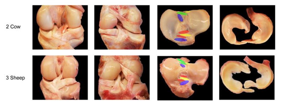
Benedikt Proffen et al. in the article titled “A Comparative Anatomical Study of the Human Knee and Six Animal Species” discuss the need for effective treatments for intra-articular knee injuries and the importance of using large animal models for translational research in knee surgery. The study aims to systematically and quantitatively compare the anatomy of the intra-articular structures of the human knee with those of six animal species (cow, sheep, goat, dog, pig, and rabbit) to determine best-practice models for experimental knee surgery.

The study involved harvesting knees from the mentioned animal species and human cadavers. The knees were thawed, and passive range of motion was measured.

Dissections were performed to reveal the anatomy of the knee structures, including cruciate ligaments (ACL and PCL), menisci (medial and lateral), tibial plateau width, and intercondylar notch width.

Measurements were taken for length, width, and area of the structures, both directly and normalized by the tibial plateau width.

Subsequently, comparative statistical analysis was conducted to identify significant differences between the human knee and animal species.

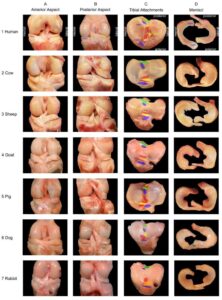
Different aspects of the knees of the seven species. [1]
Passive range of motion showed differences between quadruped animals and the human knee.The study found differences in the size and shape of the anterior cruciate ligament (ACL), posterior cruciate ligament (PCL), and menisci between species.Quantitative differences were observed in ACL and PCL dimensions between species, with variations in length and width.Tibial index, which normalizes measurements by tibial plateau width, showed variations in certain dimensions.Notch width, medial meniscus, and lateral meniscus dimensions varied between species.Anatomical differences were observed in the attachments and origins of ligaments and menisci in different species.Some animal models closely resembled specific aspects of human knee anatomy.

The study highlights the significance of choosing appropriate animal models for knee surgery research.Differences in passive range of motion between quadruped animals and humans need to be considered.Various animal models have distinct anatomical characteristics, making certain species more suitable for specific aspects of knee research.

The study provides insights into the size, shape, and anatomy of knee structures across human and animal species.

To conclude, different animal models may be suitable for various types of knee surgery research based on anatomical similarities to the human knee.

Learn More:

Proffen, Benedikt L., Megan McElfresh, Braden C. Fleming, and Martha M. Murray. “A comparative anatomical study of the human knee and six animal species.The Knee 19, no. 4 (2012): 493-499.


This article was written by Elisa Bissacco as part of an ongoing series of scientific communications written and curated by BioTrib’s Early Stage Researchers.

She is studying a PhD in Tribological Characteristics of Nanofibrous Electrospun Materials at ETH Zurich.
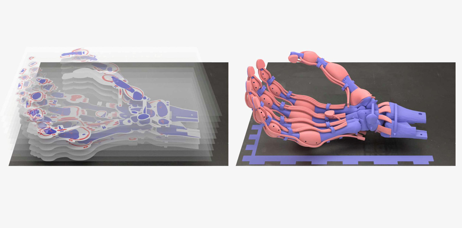
Point of Care 3D Printing

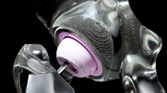
Header Image: A 3D printed acetabulum. Credit Arcam

The field of medical engineering is undergoing a transformative shift with the integration of 3D printing technology into point-of-care orthopaedics. This innovative approach has the potential to revolutionize orthopaedic treatments by reducing lead times, enhancing device fit, and minimizing material waste.

Point of care refers to the location or setting where medical diagnostics, treatments, and interventions are provided directly to patients, often in immediate proximity to the patient, facilitating rapid and convenient healthcare delivery.

Benefits of 3D Printing in Orthopaedics:
3D printing is poised to address long-standing challenges in orthopaedics, offering a fresh perspective on patient care. 3D printing introduces a sustainable approach to orthopaedics by minimizing material waste. Unlike traditional manufacturing methods, which often result in excess material usage, 3D printing enables precise fabrication, contributing to both cost savings and environmentally friendly practices (1).

Additionally, by enabling the on-site printing of bespoke medical components, 3D printing unlocks a new level of personalization, potentially resulting in improved patient outcomes (1).

This technology, that can be based all on site, has the capacity to significantly reduce lead times for patient with unique requirements where standard devices are not appropriate, which is of paramount importance, particularly in urgent and emergency situations.

Challenges and Considerations:
While the potential benefits of 3D printing in orthopaedics are immense, challenges persist. Mechanical losses during the 3D printing process can impact the durability and mechanical properties of orthopaedic devices (2). Furthermore, questions arise about the performance of 3D printed devices in tribological environments. Addressing these challenges requires ongoing research and development to optimize the technology for orthopaedic applications.

Future Possibilities:
The integration of 3D printing into point-of-care orthopaedics paints an exciting picture for the future. The ability to create personalized orthopaedic solutions on site holds promise for reducing lead times, minimizing material waste, and enhancing patient-specific implant designs. This technology has the potential to reshape the landscape of orthopaedic care and contribute to more efficient and patient-centred treatments.

The convergence of 3D printing and orthopaedics signifies a paradigm shift in medical engineering. The ability to print bespoke medical devices directly at the point of care offers a glimpse into a future where orthopaedic treatments are more tailored, efficient, and sustainable. As we navigate the challenges and opportunities presented by 3D printing, it is clear that this technology has the power to redefine the way we approach orthopaedic care, ultimately leading to improved patient outcomes and a more responsive healthcare system.


References:

[1] Teo, A.Q.A., Ng, D.Q.K., Peng, L.E.E. and O’NEILL, G.K., 2021. Point-of-Care 3D printing: A feasibility study of using 3D printing for orthopaedic trauma. Injury52(11), pp.3286-3292.

[2] Bastawrous, S., Wu, L., Liacouras, P.C., Levin, D.B., Ahmed, M.T., Strzelecki, B., Amendola, M.F., Lee, J.T., Coburn, J. and Ripley, B., 2022. Establishing 3D printing at the point of care: basic principles and tools for success. Radiographics42(2), pp.451-468.


This article was written by Ben Clegg part of an ongoing series of scientific communications written and curated by BioTrib’s Early Stage Researchers.

Ben is researching the Wear particle characterization and bio-compatibility of newly 3D printed self-lubricating polymer composites in total joint replacements at Luleå University of Technology, Sweden.

Dual effect of 3D printing Femoral Stems

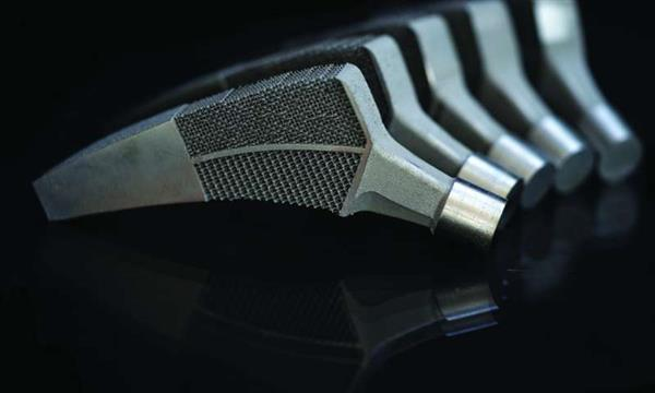
Header Image: credit to Ntop, and TU Delft


In the ever-evolving landscape of medical advancements, the marriage of 3D printing technology and topology optimization has unlocked remarkable potential in the realm of hip implants. This dynamic duo not only promises enhanced osseointegration but also addresses a critical concern – stress shielding. In this article, we delve into the groundbreaking innovation of reduced stress shielding through topologically optimized hip implants, a feat that’s transforming the way we approach orthopaedic care.

Understanding Stress Shielding

Picture this: A metal implant is introduced into a patient’s body to replace or support a damaged hip joint. Over time, the implant takes on the mechanical stresses that would naturally be borne by the surrounding bone. While this may seem like a solution, it triggers a phenomenon known as stress shielding. As the implant shoulders the load, the bone experiences reduced stress, causing it to lose density and strength. This weakened bone becomes susceptible to fractures and dislocations, negating the intended benefits of the implant.

The Perfect Harmony: Topological Optimization and 3D Printing

Enter topological optimization, a design approach rooted in mathematics that seeks to distribute stresses and strains efficiently within a structure. In the realm of hip implants, this technique takes on a revolutionary role. Combining topological optimization with the precision of 3D printing allows medical device manufacturers to craft implants that not only match a patient’s bone stiffness and density but also counteract stress shielding.

A shining example of this synergy comes from the tech prowess of IT company Altair. By harnessing the power of topological optimization software, Altair embarked on a quest to redesign the conventional hip implant.

Crafting the Future: A 3D printed Topologically-Optimized Hip Implant

Imagine a hip implant created not as a standardized component but as a bespoke masterpiece, uniquely tailored to an individual’s requirements. Armed with parameters like size, weight, and anticipated load-bearing capacity, topology optimization software worked its magic. The result? A hip implant design that ingeniously distributes stress and strain, mimicking the body’s natural mechanics.

But that’s not all. The application of topology optimization extended further, revealing areas where material could be substituted with intricate lattice structures. This strategic modification not only lightened the implant but also elevated its structural integrity.

The Game-Changing Results

The true testament of any innovation lies in its real-world impact. In one case, the topologically-optimized hip implant demonstrated a staggering 50.7% reduction in stress shielding (1). This means that the implant’s presence didn’t compromise the bone’s strength. Additionally, the implant exhibited an endurance limit that could endure a jaw-dropping 10 million cycles – equivalent to jogging from Los Angeles to New York and back, not just once but twice!

Challenges

The path to innovation is seldom without obstacles. As we celebrate the potential of topologically-optimized hip implants, we must also acknowledge the challenges that lie ahead.

  1. Sterilization Difficulties: 3D-printed implants introduce new considerations in sterilization. The intricate lattice structures that contribute to implant performance may also harbor contaminants, necessitating novel sterilization techniques that preserve the implant’s structural integrity.
  2. Corrosion Concerns: The unique geometries and materials used in 3D printing can introduce corrosion risks. Addressing this challenge requires the development of corrosion-resistant materials and surface treatments to ensure the implant’s long-term viability.

A Glimpse into the Future

If we can overcome these challenges, the fusion of topological optimization and 3D printing has bestowed the medical world with a transformative tool. As we witness stress shielding dissipate before our eyes, we catch a glimpse of the future – one where hip implants, and potentially other medical devices, become personalized marvels, engineered to harmonize seamlessly with the human body.

In closing, the convergence of mathematical ingenuity and cutting-edge technology is sculpting a path towards a new era of orthopaedic care. Reduced stress shielding is no longer a distant dream; it’s a reality that holds the promise of stronger, healthier tomorrows for patients around the world.


Learn More:

[1]

7 Complex Designs Achieved With 3D Printing


This article was written by Ben Clegg part of an ongoing series of scientific communications written and curated by BioTrib’s Early Stage Researchers.

Ben is researching the Wear particle characterization and bio-compatibility of newly 3D printed self-lubricating polymer composites in total joint replacements at Luleå University of Technology, Sweden.

The raise of biodegradable implants for fracture fixation devices

Biodegradable implants for bone fracture fixation are a relatively new technology that has the potential to revolutionize the way we treat bone fractures. These implants are made from biodegradable materials that are designed to dissolve over time, eliminating the need for additional surgery to remove them.

One of the main benefits of biodegradable implants is that they can help reduce the risk of infection and complications associated with traditional metal implants. They also avoid the need for a second surgery to remove the implant, which can be costly and time-consuming.
Another advantage of biodegradable implants is that they can provide support for the bone as it heals, while also promoting the growth of new bone tissue. This can help to improve the overall healing process and reduce the risk of complications such as non-union or malunion of the bone.

There are currently several types of biodegradable implants available for use in bone fracture fixation, including those made from polylactic acid (PLA), polycaprolactone (PCL), polydioxanone (PDO) and also metal based alternatives. Each of these materials has its own unique properties that make it suitable for different types of fractures and patients. Among the metal ones a lot of interest is raising around magnesium alloys that have the advantage of having appropriate mechanical properties, high biocompatibility and to promote bone in-growth.

It is important to note that biodegradable implants are not suitable for all types of fractures or patients. They are typically used for fractures that are considered to be low-risk and are not expected to experience high levels of stress during the healing process.

Despite these limitations, biodegradable implants are a promising technology that could have a significant impact on the way we treat bone fractures in the future. As research in this area continues to advance, we can expect to see even more innovative and effective biodegradable implant options for patients.

In conclusion, biodegradable implants for bone fracture fixation are a relatively new technology that holds great promise for the future. These implants are made from biodegradable materials that are designed to dissolve over time, eliminating the need for additional surgery to remove them. They can help reduce the risk of infection and complications associated with traditional metal implants, while promoting the growth of new bone tissue. With more research, the limitations of these implants can be overcome and they can be used to treat a wider range of fractures.


References:

[1] K. Kumar, R. S. Gill, and U. Batra, “Challenges and opportunities for biodegradable magnesium alloy implants,” Mater. Technol., vol. 33, no. 2, pp. 153–172, Jan. 2018, doi: 10.1080/10667857.2017.1377973.


This article was written by Giulio Cavaliere as part of a series articles curated by BioTrib’s Early Stage Researchers.

Giulio Cavaliere is investigating Additively manufactured biodegradable alloys for bone replacement at Uppsala University, Sweden.

Valborg: a Swedish tradition

Valborg, also known as Walpurgis Night, is an annual Swedish tradition that takes place on the last day of April. This celebration marks the arrival of spring and is a time for people to come together and celebrate the end of winter.Valborg is a particularly special tradition in the city of Uppsala, located in central Sweden. The city is home to one of the oldest universities in Sweden, Uppsala University, and the celebration of Valborg is particularly grand and lively in the city, with a festive atmosphere and many different events taking place.

The origins of Valborg can be traced back to the pre-Christian era, when the ancient Germanic people celebrated the arrival of spring with a festival called Walpurgis Night. The name “Valborg” is derived from the name of the Christian saint Walpurga, who was celebrated on May 1st in medieval times.

The city of Uppsala also has a tradition of boat parades on the Fyris river, where people gather to watch beautifully decorated boats passing by, with music and singing. The more than 100 polystyrene boats are carved and decorated by students and compete to survive the longest through a series of small waterfalls.
Another popular event is the Valborg concert held at the Uppsala Cathedral, featuring choirs and brass bands.


This article was written by Giulio Cavaliere as part of a series articles curated by BioTrib’s Early Stage Researchers.

Giulio Cavaliere is investigating Additively manufactured biodegradable alloys for bone replacement at Uppsala University, Sweden.

The Benefits and Risks of Hip Replacement Surgery

Hip replacement surgery, also known as hip arthroplasty, is a common procedure that can relieve pain and improve mobility in individuals with hip joint damage. The procedure involves replacing the damaged joint with an artificial one, typically made of metal or plastic. The benefits of hip replacement surgery include a significant reduction in pain, increased mobility and an improved quality of life. Many people who have hip replacement surgery report that their pain is greatly reduced, and their ability to perform daily activities is improved.

One of the most common reasons for hip replacement surgery is osteoarthritis, which is a degenerative joint disease that causes the cartilage in the joint to wear away. This can result in bone-on-bone contact, causing pain and stiffness. Other reasons for hip replacement surgery include rheumatoid arthritis, avascular necrosis (death of bone tissue due to lack of blood supply) and fractures.

Hip replacement surgery is typically considered when other treatments, such as physical therapy, medication, and lifestyle changes, have failed to provide relief. The procedure is typically performed under general anaesthesia and can take several hours to complete. After the surgery, patients will need to go through a period of recovery and rehabilitation to help them regain their mobility and strength.

It is important to note that like any surgical procedure, there are risks involved such as infection, bleeding, blood clots and implant failure. In some cases, the implant may not function properly or may become loose, requiring a revision surgery. Additionally, there is a risk of nerve or blood vessel injury during the procedure.

However, the success rate for hip replacement surgery is quite high and most people who have the procedure experience a significant improvement in their pain and mobility. It is important to discuss the potential risks and benefits of the surgery with your doctor to determine if it is the right choice for you.


This article was written by Ben Clegg part of an ongoing series of scientific communications written and curated by BioTrib’s Early Stage Researchers.

Ben is researching the Wear particle characterization and bio-compatibility of newly 3D printed self-lubricating polymer composites in total joint replacements at Luleå University of Technology, Sweden.


Pros and Cons of various Total Hip replacements

Total hip replacement (THR) is a common procedure used to relieve pain and improve mobility for those suffering from hip arthritis or other hip-related conditions. However, there are different types of THR available, each with their own pros and cons.

One type of THR is known as a cemented hip replacement, usually for older patients with less remaining healthy bone around the femur and acetabular components. The cement helps secure the implant into place along with the slightly weaker bone. This procedure involves using a cement to secure the prosthetic implant to the natural bone. Pros of this procedure include a high success rate and the ability to return to normal activities quickly. Cons include a longer recovery time and a higher risk of complications.

Another type of THR is known as an uncemented hip replacement. Usually for the younger profile of patients, that have healthy bone around the hip, that is able to regrow and secure itself onto the surface of the implant, ensuring a symbiotic relationship between the human and the implant. This procedure does not use cement, instead, the implant is designed to bond with the natural bone over time. Pros of this procedure include a lower risk of complications and a shorter recovery time. Cons include a slightly lower success rate and a longer rehabilitation period.

A third type of THR is called a hybrid hip replacement. This is usually for patients that have healthy bone, but a section of the femur or acetabular has been damaged or compromised (via disease or physical impact) so that that area needs some cement to help secure the implant. This procedure involves using a combination of cement and uncemented techniques. Pros of this procedure include a high success rate and a shorter recovery time. Cons include a higher risk of complications and a longer rehabilitation period.

In conclusion, the type of total hip replacement you choose will depend on your individual needs and preferences. It is important to discuss the pros and cons of each type with your surgeon to determine the best option for you. Ultimately, the goal of any hip replacement is to relieve pain and improve mobility, so it is important to choose the option that will best achieve that goal for you.


This article was written by Ben Clegg part of an ongoing series of scientific communications written and curated by BioTrib’s Early Stage Researchers.

Ben is researching the Wear particle characterization and bio-compatibility of newly 3D printed self-lubricating polymer composites in total joint replacements at Luleå University of Technology, Sweden.

Hip Replacement Surgery: What to Expect Before, During, and After

Before the surgery, the patient will undergo pre-operative tests and evaluations to ensure that they are fit for the procedure. This may include blood tests, an EKG, and X-rays of the hip joint. The patient will also meet with the anaesthesiologist to discuss the type of anaesthesia that will be used during the procedure.

During the surgery, the damaged joint will be removed and replaced with an artificial one. The procedure is typically performed under general anaesthesia and can take several hours to complete. The surgeon will make an incision in the hip and remove the damaged joint. The new joint will then be positioned and secured in place.

After the surgery, patients will need to go through a period of recovery and rehabilitation to help them regain their mobility and strength. This may include physical therapy, pain management, and the use of assistive devices such as crutches or a walker. The recovery time and success of the surgery may vary depending on the individual’s overall health and the type of implant used.

In the first few weeks after surgery, patients will be advised to limit their activity and avoid putting weight on the affected hip. As the hip begins to heal, patients will gradually increase their activity level and begin to regain their strength and mobility. The length of the recovery period can vary from person to person, but most people are able to return to their normal activities within a few months.

It is important to note that hip replacement surgery is not a cure for the underlying condition that caused the joint damage. However, it can greatly improve the patient’s quality of life by reducing pain and improving mobility.


This article was written by Ben Clegg part of an ongoing series of scientific communications written and curated by BioTrib’s Early Stage Researchers.

Ben is researching the Wear particle characterization and bio-compatibility of newly 3D printed self-lubricating polymer composites in total joint replacements at Luleå University of Technology, Sweden.


MC3T3 cells: Choose your media

If you work with in vitro bone analysis, probably you have already used the pre-osteoblastic cell line called MC3T3-E1. Nowadays, this is the most used cell line for bone focused experiments. This cell line was selected amongst several subclones because it showed a high differentiation rate (cell commitment to a cell line) and mineralization (calcium deposition) after being grown in media containing ascorbic acid. Of course, it also can be used for other testing e.g., proliferation or cytotoxicity.

Almost all in vitro studies regarding material biocompatibility for bone regeneration use MC3T3 cells, sometimes together with other cell types, so it is really important to deeply understand their behavior. Of particular importance, as mentioned above, is the presence of vitamin C in the growth medium.

In a paper that I have recently stumbled upon [1], Izumiya et al analyze the effect of different commercial cell medium that contain vitamin C on MC3T3 cells. They also compared those with some homemade media with different amount of ascorbic acid in it. This paper shows that the use of different media can substantially modify the outcome of an experiment, regarding: cell proliferation, differentiation and mineralization.

The real reason behind this experiment is that, despite MC 3T3 cells are used worldwide and on a daily basis, there is no standardized growth protocol and each experiment is performed under laboratory-specific culture conditions [1]. In my opinion, this is leading the scientific community to possible misinterpretations of data and it is really difficult to compare data from different studies. Reading this paper or simply being aware of the problem is surely the first step to contribute to a better in vitro system analysis.


References:

[1] Izumiya et al (2021). Evaluation of MC3T3-E1 Cell Osteogenesis in Different Cell Culture Media. Molecular Science. https://doi.org/10.3390/ijms22147752.


This article was written by Niccoló De Berardinis as part of an ongoing series of scientific communications written and curated by BioTrib’s Early Stage Researchers.

Niccoló is researching Bioimaging of biomaterials and biological characterization of 3D-printed alloys for reconstructive surgery at Uppsala University, Sweden.

Mandatory Courses at Uppsala University

As many universities do, Uppsala University offers to its PhD students the possibility to include some courses into their study plan, but it also requires some mandatory courses as well to be included in it. The courses vary from department to department and, in my case, there were three of them. To be more precise, I had to take an: ethic, biostatistics and a scientific presentations course.

To be honest, I started this mandatory process thinking if that was really necessary since we all went through a really hard PhD selection and also a MSc and BSc studies in which we have already demonstrated our knowledge and skills (in my case also in the course contents). So, to put it bluntly, I was not very thrilled at the idea, but of course I understand the thought behind it and objectively speaking these are really important and necessary things to know, so it makes sense that a great institution such as Uppsala University want to be sure that its students are very well acquainted with these arguments. We are talking about standards here, and for sure it is good to have them both in science and in other fields.

But, when I really started, since the very first lessons of the first course, I truly understood that I was wrong. Of course, they were talking about things that I already know, but they were also putting us in the right state of mind and disposition to confront possible problems that we could find in our future career. Now I have finally took all three courses and I really can say that I was definitely enriched by them. I am with a much wider network, knowledge and understandings. So, I guess that the morale here is, never get something for granted even if you think that you know everything about it. Sometimes another perspective can give you the insights that you were searching for, especially in science.


This article was written by Niccoló De Berardinis as part of an ongoing series of scientific communications written and curated by BioTrib’s Early Stage Researchers.

Niccoló is researching Bioimaging of biomaterials and biological characterization of 3D-printed alloys for reconstructive surgery at Uppsala University, Sweden.

Bone Analysis Guide

Undoubtedly, choosing the right set-up for your experiment is one of the most critical steps when working in science. The problems that we scientists want to solve, the innovation that we want to bring, are almost always very complex phenomena. This is why we have to simplify them into small experiments from which we can build our thesis.

My research is based on biomaterials developed for orthopedic purposes, therefore, I work a lot with bone cells, bone related assays, and whatnot. So, if you are searching for a handy guide on how to evaluate osteoimmunomodulatory properties of biomaterial you have come to the right place. I always suggest to my student to read and fully understand a review article by Mestres et al [1], before starting to do some lab work. The title of the paper is: “A practical guide for evaluating the osteoimmunomodulatory properties of biomaterials”. In this case, I know personally some of the authors so perhaps I am a little bit biased, but I will let you decide should you want to go through it.

In my opinion, this paper contains all the information needed to someone that is trying to approach to the bone-biomaterials world. It contains background and advanced knowledge about the interaction between immune cells and bone cells, the fracture healing and the bone remodelling processes and also a lot of information about cell types, methodologies and cellular assays to run in vitro testing on your biomaterial.

So should you want to change your in vitro experimental approach, take a look at this amazing work.


Header Image: Graphical Abstract of A practical guide for evaluating the osteoimmunomodulatory properties of biomaterials [1].


References:

[1] Mestres et al (2022). A practical guide for evaluating the osteoimmunomodulatory properties of biomaterials. Acta Biomaterialia. https://doi.org/10.1016/j.actbio.2021.05.038.


This article was written by Niccoló De Berardinis as part of an ongoing series of scientific communications written and curated by BioTrib’s Early Stage Researchers.

Niccoló is researching Bioimaging of biomaterials and biological characterization of 3D-printed alloys for reconstructive surgery at Uppsala University, Sweden.

The importance of learning the local language

Header Image Source: The Best Way To Learn German: What You Probably Don’t Know (bestplacestovisitgermany.com)

Multilingualism has evolved beyond being merely “essential” in the modern world. It has become clear that learning a language different than your mother tongue is quite advantageous. For example, for an international PhD student like me, learning German in Switzerland is highly important for a variety of reasons. Switzerland is a multilingual country with four official languages: German, French, Italian and Romansh. German is the most widely spoken language in Switzerland and is used in many official and business contexts. Knowing German can be especially important for those living in the German-speaking parts of Switzerland, such as Zurich and Bern.

First, being able to speak German in Switzerland can greatly improve one’s ability to communicate with the local population, including in the workplace. It allows for better integration and inclusion into Swiss society, making it easier to form connections and build relationships with colleagues and friends.

Additionally, having a good command of German can open up many more opportunities for employment in Switzerland. Many Swiss companies conduct business in German, and many jobs require German language skills. Furthermore, German is often used as a common language in international companies and organizations based in Switzerland, making it a valuable skill for those looking to work in such environments.

Furthermore, Understanding and being able to speak German also allows for greater access to Swiss culture and society. German is the language of many newspapers, books, and other forms of media in Switzerland, and understanding the language can help to better understand the country’s culture, history, and politics.

In summary, learning German in Switzerland is important for inclusion, communication, working opportunities and cultural understanding. Having a good command of German can greatly improve one’s ability to navigate and succeed in Swiss society, whether in a personal or professional context.

“Auf Wiedersehen!”


This article was written by Alessio Amicone as part of an ongoing series of scientific communications written and curated by BioTrib’s Early Stage Researchers.

Alessio is investigating the Elucidation of Friction-Induced Failure Mechanisms in Fibrous Collagenous Tissues at ETH Zürich, Switzerland.