Peter Jost, the father of tribology

We always talk about tribology and biotribology, after all, it is our main topic in this fantastic project. But to whom do we owe the birth of tribology? During the network-wide training being held in London, Connor Myant started the conference by asking: “do you know who Peter Jost was? The man to whom we owe the birth of tribology”. But why is Peter Jost considered the father of tribology?

Only after eye-opening British research called “The Jost Report” in the 1960s did tribology start to receive widespread recognition. In fact, in 1964, the UK Department of Education and Science asked a working committee led by Dr. H. Peter Jost to look at the condition of lubrication education and research and to offer their judgment on the requirements of industry.

Cover of the Jost Report [1]
When the group reported in February 1966, it proposed a new word to describe this multidisciplinary field – tribology, from the Greek root “τριβo” meaning rubbing or attrition [1]. According to the Oxford English Dictionary tribology is defined as “the branch of science and technology concerned with interacting surfaces in relative motion and with associated matters (as friction, wear, lubrication, and the design of bearings)”.

Mechanical engineers, materials scientists, physicists, and chemists were needed to advance tribology. In consequence, tribological developments have supported a large portion of engineering development worldwide. Tribology is most frequently linked with bearing design, but it has applications in all sectors of contemporary technology where surfaces interact, including seemingly unexpected ones like hair conditioners and cosmetics [1].

Jost’s work in tribology has been instrumental in establishing the field as it is today. An output from this Jost’s reviews was the development of the field of biotribology – Championed by Prof Dowson at the University of Leeds. Biotribology is the subfield of tribology which deals with the interactions between biological systems and their environment, specifically the friction, wear, and lubrication of biological systems. Prof Dowson’s contributions to establishing accepted lubrication equations and drive innovation in testing and materials tribology has helped to improve the design and function of medical devices, implants, and prostheses, and have had a significant impact on the fields of biomechanics, biomaterials, and biomedicine.

Jost passed away on June 2016, but his legacy lives on through the countless researchers, engineers, and practitioners who have been influenced by his work.

References

1-Fifty years of tribology | Department of Engineering (cam.ac.uk)


This article was written by Alessio Amicone as part of an ongoing series of scientific communications written and curated by BioTrib’s Early Stage Researchers.

Alessio is investigating the Elucidation of Friction-Induced Failure Mechanisms in Fibrous Collagenous Tissues at ETH Zürich, Switzerland.

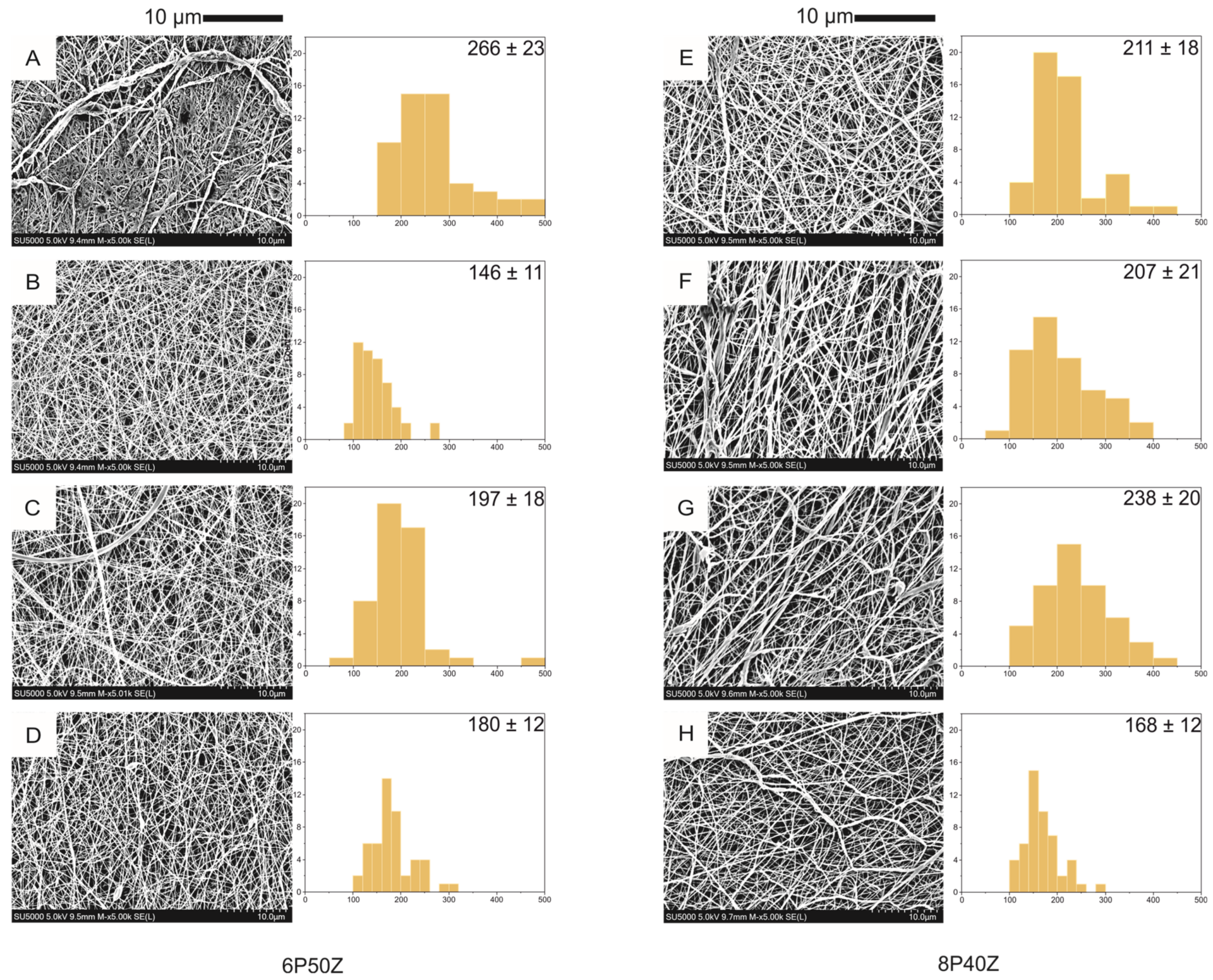
Published paper: Co-electrospun PCL/Zein membranes as articular cartilage engineering scaffolds

Header image: Nanofiber morphology of PCL/zein electrospun mats prepared from a 70/30 vol./vol.

A recent publication titled “Co-electrospun PCL/Zein membranes as scaffolds for articular cartilage engineering” by the ETH team was featured in MDPI Bioengineering. This paper builds upon the research initially presented at ETH’s Materials and Processes seminar in 2022. The study explores the application of Zein, a corn protein widely used in food packaging and drug encapsulation industries, in the development of tissue engineering scaffolds for articular cartilage.

Through co-electrospinning, the team successfully reduced protein adsorption by half compared to scaffolds made solely of bovine serum albumin and equine synovial fluid. Additionally, the PCL/Zein membranes exhibited lower roughness when compared to pure PCL scaffolds. These favorable characteristics are believed to be attributed to the enhanced spreading of bovine chondrocytes cultivated on the membrane surfaces.

Overall, this research highlights the potential of co-electrospun PCL/Zein membranes as promising scaffolds for articular cartilage engineering, showcasing their improved protein adsorption properties and reduced surface roughness.

Check out the paper below:

Plath, A.M.S.; Huber, S.; Alfarano, S.R.; Abbott, D.F.; Hu, M.; Mougel, V.; Isa, L.; Ferguson, S.J. Co-Electrospun Poly(ε-Caprolactone)/Zein Articular Cartilage Scaffolds. Bioengineering 202310, 771. https://doi.org/10.3390/bioengineering10070771


This article was written by André Plath as part of a series articles curated by BioTrib’s Early Stage Researchers.

André is one of BioTrib’s Early Stage Researcher‘s who is investigating Boundary Lubrication of Fibrous Scaffolds at ETH Zürich, Switzerland.

Celebrating Pride Month: Championing Diversity in Medical Engineering

As the colorful and vibrant month of June rolls around, we find ourselves immersed in the spirit of LGBTQIA Pride Month, a time to honor and celebrate the diverse identities within the queer community. While Pride Month resonates with people from all walks of life, it is particularly significant for researchers and academics in STEM, including the field of medical engineering. This article delves into the importance of LGBTQIA representation in the realm of medical engineering and highlights the strides made by the community.

Promoting Inclusivity in Medical Engineering:

Inclusivity is the cornerstone of progress in any field, and medical engineering is no exception. By embracing diverse perspectives and fostering an inclusive environment, we encourage creativity, innovation, and breakthroughs. LGBTQIA researchers and academics bring unique insights and experiences that can profoundly impact medical engineering’s trajectory.

Supporting LGBTQIA Students and Professionals:

Creating a welcoming space for LGBTQIA individuals in medical engineering is paramount. Institutions and organizations can play a vital role by establishing LGBTQIA support networks, mentorship programs, and providing resources for queer students and professionals. Recognizing the specific challenges faced by LGBTQIA individuals in the workplace and academia, such initiatives can foster a sense of belonging and help overcome barriers.

Research on LGBTQIA Health:

While tremendous progress has been made, significant gaps remain in understanding and addressing LGBTQIA health disparities. Medical engineers have an opportunity to contribute by focusing their research on LGBTQIA-specific health issues, such as gender-affirming surgeries, hormone therapy, mental health support, and improving healthcare access. By addressing these needs, researchers can directly impact the lives of LGBTQIA individuals, promoting equitable healthcare outcomes.

Collaboration and Intersectionality:

The LGBTQIA community intersects with various other identities, and recognizing this intersectionality is crucial. Collaborative efforts between medical engineers, clinicians, psychologists, and other professionals can lead to a more comprehensive understanding of LGBTQIA health concerns and generate innovative solutions.


As we celebrate Pride Month, let us take this opportunity to recognize the importance of diversity and inclusivity in medical engineering. By fostering an environment that welcomes LGBTQIA+ researchers and professionals, supporting their unique perspectives, and focusing on LGBTQIA+ health disparities, we can make significant strides towards a more equitable and inclusive future for all. Together, let us champion diversity and create a world where every individual’s identity is celebrated and valued. Happy Pride Month!

Inclusive Language: Beyond the political correctness

On 26 May 2023, BioTrib hosted Tatiana Vieira, director of the Brasilea Foundation in Switzerland. Her talk “Inclusive Language: Beyond the Political Correctness” was watched by consortium members. For one hour, the participants practices to avoid sexist, racist, and other biased or prejudiced ideas were discussed. Tatiana also shared her experiences presenting two case studies at the SWI and Radio X a radio and tv broadcast.

In summary, inclusive language consists of practices to eliminate noise in communication and avoid discrimination and the feeling of exclusion based on gender identity, ethnicity, age, sexual orientation, etc.  In an international and diverse group, this is an important debate. With the talk, we expect to incorporate inclusive language into our communications and make the network more inclusive.


This article was written by André Plath as part of a series articles curated by BioTrib’s Early Stage Researchers.

André is one of BioTrib’s Early Stage Researcher‘s who is investigating Boundary Lubrication of Fibrous Scaffolds at ETH Zürich, Switzerland.

The importance of resilience in graduate school

Research is messy and full of failed attempts. Trying to protect students from that reality does them a disservice.

In “Why I teach my students about scientific failure”, Jennifer Lanni discusses the experience of giving students failed western blot results and the discussions these triggered. The students’ deception of not having precise results stimulates deep discussions and potential troubleshooting routes.

The text resonated with some of the feelings I always had. When presenting seminars and in group meetings, I frequently felt I hid some of the mishaps in my project. I try showing the things that worked instead of showing the process and troubleshooting behind the failed experiments. The text opened my eyes to academic honesty and how we all could benefit from showing “some weaknesses”. It also gave me a better understanding of how open data would benefit us all in the scientific community and how different groups could learn from others’ failures. I believe in a world with more dissemination and open failure communication, less data fabrication and fraud would exist.


References

Why I teach my students about scientific failure, AAAS Articles DO Group. (2021). https://doi.org/10.1126/science.caredit.acz9901.


This article was written by André Plath and Giulio Cavaliere as part of a series articles curated by BioTrib’s Early Stage Researchers.

André is one of BioTrib’s Early Stage Researcher‘s who is investigating Boundary Lubrication of Fibrous Scaffolds at ETH Zürich, Switzerland.

Are my methods addressing the research questions? Addressing methodology and reproducibility in biomaterials science

Ph.D. students, myself included, often ask themselves: Is my research leading somewhere? Am I going to create any impact with my project? The daily chores, “publish or perish”, and increasing competition for senior positions push students to create data in a record time; however, not everything that is published is reproducible or could lead to clinical/translational applications.

To address this, Daniel Lakens in a recent World View paper for nature discusses the importance of creating methodological review boards. In his opinion, by having methods scrutinized, students could approach or phrase the initial research questions more efficiently, and determine what tests should be made, sample size, etc.  Baker gives several tips on how to publicize protocols and increase reproducibility. She mentions that people might work with the same materials and obtain different results, and for eliminating this, thorough descriptions and revisions by peers are necessary. For this, she lists a series of online tools.

These two cases might not be the solution to all problems, but they certainly address some of them. By reviewing the methodology and aligning it to the initial research question, early-career scientists might make progress faster and have their daily struggles reduced.

References

Lakens, D. (2023) Is my study useless? why researchers need methodological review boards, Nature News. Available at: https://www.nature.com/articles/d41586-022-04504-8 (Accessed: 24 May 2023).

Baker, M. (2021) Five keys to writing a reproducible lab protocol, Nature News. Available at: https://www.nature.com/articles/d41586-021-02428-3 (Accessed: 24 May 2023).


This article was written by André Plath and Giulio Cavaliere as part of a series articles curated by BioTrib’s Early Stage Researchers.

André is one of BioTrib’s Early Stage Researcher‘s who is investigating Boundary Lubrication of Fibrous Scaffolds at ETH Zürich, Switzerland.

Surface texturing of Ti-6Al-4V alloy towards improvements in corrosion, tribocorrosion and biocompatibility properties

Ti-6Al-4V alloy is widely used in aircraft, automotive and biomedical applications due to its corrosion resistance, high strength-to-weight ratio (i.e., specific strength) and biocompatibility properties. Even though these characteristics are required in metal components used in total joint replacement surgeries, Ti-6Al-4V exhibits a poor tribological performance.

Different post-processing approaches (e.g., heat treatments, surface coating, and surface texturing) have been investigated to tackle this drawback. Laser texturing, for instance, has become an increasingly post-processing route for improving the corrosion resistance, tribological behaviour and biocompatibility of Ti-6Al-4V surfaces. In the work of Wang and collaborators (2022), they investigated those properties by creating a microgrooved surface on the alloy via UV nanosecond laser texturing.

An enhancement in corrosion resistance was found in laser texture surfaces, which might be due to a β → α phase transformation occurring in the surface motivated by laser ablation. On a similar note, the tribological performance of the surface treated material displayed an enhancement (i.e., reduction of coefficient of friction during dry sliding and decrease in wear volume generated). The authors attribute this phenomena to an augmentation in the surface hardness of the material also caused by laser texturing.

In vitro bioactivity, evaluated via BMSC adhesion, also followed the trend of the before-mentioned properties, with microgrooved surfaces showing the highest proliferation rate and adhesion number.

Header image reproduced from Wang (2022).

References:

Wang, C., Tian, P., Cao, H., Sun, B., Yan, J., Xue, Y., … & Zhao, X. (2022). Enhanced Biotribological and Anticorrosion Properties and Bioactivity of Ti6Al4V Alloys with Laser Texturing. ACS omega, 7(35), 31081-31097.


This post was written by Pedro Luiz Lima dos Santos as part of an ongoing series of scientific communications written and curated by BioTrib’s Early Stage Researchers.

Pedro is researching the Functional Biotribology of the Surface Engineering of 3D Printed Components at the University of Leeds, UK.

Biotribology in nature: how different microstructure morphologies change leaf wettability?

Header Image: The surface morphology with the magnification of one thousand times of the four kinds of plant leaves: (a) Photinia serrulata, (b) Ginkgo, (c) Aloe vera and (d) Hypericum monogynum. (CC BY-NC-ND 4.0)

Throughout millions of years, organisms evolved in Nature due to a need of adaptation driven by different environmental conditions imposed. Functional systems with intricate properties arose from this continuous structural development leading to, for example, super hydrophobic and self-cleaning surfaces found in lotus leafs (referred as “lotus effect”). An understanding of role of the microstructural features in these systems may help elucidating how to tailor system with an appropriate surface wettability.

In order to tackle this need, Wang and co-workers (2016) studied the wettability properties of four different types of plants (P. serrulata, Ginkgo, Aloe vera, H. monogynum) exhibiting dissimilar microstructures by means of static contact angle for deionized water.

Their results provide an insightful understanding of surface wettability. Whilst minor corrugated and raised boundary microstructures portray the highest wettability (i.e., P. serrulata), increase in cross section corrugation diminishes the liquid/surface contact area and, therefore, intensifies hydrophobicity. Also, the L/W ratio seems to play a major role in surface wettability. Ginko, although displays a corrugated microstructure on the leaf, its large L/W ratio promotes diffusion of liquid, which consequently leads to a hydrophilic surface.

References:

Wang, L. F., & Dai, Z. D. (2016). Effects of the natural microstructures on the wettability of leaf surfaces. Biosurface and Biotribology, 2(2), 70-74.

This post was written by Pedro Luiz Lima dos Santos as part of an ongoing series of scientific communications written and curated by BioTrib’s Early Stage Researchers.

Pedro is researching the Functional Biotribology of the Surface Engineering of 3D Printed Components at the University of Leeds, UK.

Tissue Engineering for Articular Cartilage Regeneration

Light micrograph of hyaline cartilage

Articular cartilage is a living material composed of a relatively small number of cells known as chondrocytes surrounded by a multicomponent matrix. Mechanically, articular cartilage is a composite of materials with widely differing properties. Approximately 70 to 85% of the weight of the whole tissue is water. The remainder of the tissue is composed primarily of proteoglycans and collagen. Proteoglycans consist of a protein core to which glycosaminoglycans (chondroitin sulfate and keratan sulfate) are attached to form a bottlebrush-like structure.

The structure of articular cartilage is often described in terms of four zones between the articular surface and the subchondral bone: the surface or superficial tangential zone, the intermediate or middle zone, the deep or radiate zone, and the calcified zone.

Currently, the most used techniques for articular cartilage regeneration are microfracture (MF), osteochondral autologous transplantation (OAT), osteochondral allograft transplantation (OCA), particulate articular cartilage implantation (PACI), and autologous chondrocyte implantation (ACI). However, these methods have limitations including calcification, formation of transient fibrocartilaginous tissue, and the low capacity of binding to surrounding normal cartilage [1, 2]. Therefore, scaffolds with improved bulk mechanical properties could increase the efficacy of treatment and promote an earlier return to normal activity.

In recent years, tissue engineering technology has been considered the most promising method for regenerating the articular cartilage [3] [4].

In tissue engineering applications, biomaterial scaffolds play animportant role in providing a 3D environment that supports cellgrowth, matrix deposition, and tissue regeneration. An ideal tissue engineering scaffold should meet several important criteria:

  1. Be biocompatible, minimizing local tissue reactions andmaximizing cell growth and tissue integration.
  2. Be biodegradable with good absorption rate, providing support for early cell proliferation and allows for gradualdegradation after the formation of new tissue.
  3. Have adequate porosity and interconnectivity to allow cellmigration and efficient exchange of nutrients and waste.
  4. Possess suitable mechanical properties to support tissue growthunder natural mechanical loads.


To date, many biomaterial scaffolds have been extensively studied, including natural polymers extracted from living organisms and synthetic materials derived from various chemical processes used intissue repair and regeneration.

Natural biomaterials are popular as scaffolds for cartilage repair and regeneration due to their excellent biocompatibility for cell adhesion and differentiation. In particular, natural scaffolds used in tissue engineering of articular cartilage include carbohydrate-based hyaluronic acid, agarose, alginate, chitosan, and protein-based collagen or fibrin glues.

Due to its ease of fabrication and chemical modification, excellent biocompatibility, high versatility, suitable mechanical properties and controllable biodegradability, synthetic polymers are currently being investigated for their potential as a scaffold for cartilage tissue. The most common synthetic polymers for cartilage engineering scaffolds are polylactic acid (PLA, present in both L and D forms), polyglycolicacid (PGA), and its copolymer poly-lactic-co- Glycolic acid (PLGA).

Conventional natural or synthetic scaffolds still need to be improved to achieve better biocompatibility and functional properties for cartilage regeneration. Because the size of native cartilage tissue is only nanometers, and chondrocytes directly interact with nanostructured ECM, the biomimetic properties and excellent physicochemical properties of nanomaterials are essential for chondrocyte growth. [5].

Solution electrospinning (SES) is a technique that allows the production of nanofibrous scaffolds and allows for the tuning of the 3D scaffolds by changing the fiber diameter and scaffold porosity. The process consists of a pump that pushes out, through a metal needle (spinneret), the polymer solution, inserted in a syringe. The presence of a high voltage source that energizes the polymer solution causes formation of a conical jet (Taylor cone) which is then drawn into a fiber by electrostatic repulsion [5] [6]. The resulting fibers are deposited on a flat or tubular electrode (collector). The thin electrospun fibers range from a few hundred nanometers to a few micrometers and are suitable candidates to mimic the structure of the natural extracellular matrix (ECM) as they can stimulate cell ingrowth and proliferation [7].

Advances in fabrication methods have solved the scalability problem and enabled the development of porous structures that allow long-term cell invasion and growth. Despite all these advantages, electrospun scaffolds have yet to be fully evaluated in preclinical models and clinical settings, hindering widespread acceptance of this breakthrough technology in biomedicine [8]

Bibliography

[1] C. Vinatier and J. Guicheux, “Cartilage tissue engineering: From biomaterials and stem cells to osteoarthritis treatments,” Annals of Physical and Rehabilitation Medicine, vol. 59, pp. 139 – 144, 2016.

[2] W. Wei, Y. Maa, X. Yao, W. Zhou, X. Wang, L. Chenglin, J. Lin, Q. He, S. Leptihna and H. Ouyang, “Advanced hydrogels for the repair of cartilage defects and regeneration,” Bioactive Materials, vol. 6, p. 998–1011, 2013.

[3] S. Jiang, W. Guo, G. Tian, X. Luo, L. Peng, S. Liu, X. Sui, Q. Guo and X. Li, “Clinical Application Status of Articular Cartilage Regeneration Techniques: Tissue-Engineered Cartilage Brings New Hope,” Stem Cells International, 2020.

[4] A. Martín, H. Zlotnick, J. Carey and R. Mauck, “Merging therapies for cartilage regeneration in currently excluded ‘red knee’ populations,” Nature Partner Journal Regenerative Medicine, vol. 4, 2019.

[5] N. Maurmann, S. L and P. P, “Electrospun and Electrosprayed Scaffolds for Tissue Engineering,” Cutting-Edge Enabling Technologies for Regenerative Medicine, pp. 79 – 100, 2018.

[6] R. Soares and al., “Electrospinning and electrospray of bio-based and natural polymers for biomaterials development,” Mater Sci Eng C Mater Biol Appl, pp. 969-982, 2018.

[7] D. Alexeev and al., “Electrospun biodegradable poly(epsilon-caprolactone) membranes for annulus fibrosus repair: Long-term material stability and mechanical competence,” JOR Spine, vol. 1, 2021.

[8] E. Z. D. Yilmaz, “Electrospun Polymers in Cartilage Engineering—State of Play,” Front. Bioeng. Biotechnol., 2020.

[9] L. H. J. A. K. Zhang, “The Role of Tissue Engineering in Articular Cartilage Repair and Regeneration,” NIH Public Access, 2009.


This article was written by Elisa Bissacco as part of an ongoing series of scientific communications written and curated by BioTrib’s Early Stage Researchers.

She is studying a PhD in Tribological Characteristics of Nanofibrous Electrospun Materials at ETH Zurich.

Implant Impressions Episode 1: Anne

Are you considering joint replacement surgery and want to hear firsthand accounts of the procedure? We recently had the pleasure of interviewing a joint replacement surgery patient (Anne, 85) to learn more about their experience and gain insight into their patient perspective. This video will offer a look into the subjective experience, thoughts and feelings of a person who has gone through the joint replacement surgery process.

Self-driven Biomedical sensor: in situ wear debris from artificial hip joint

Header image adapted from Liu, 2021. Left: Working mechanism of the fabricated TENG. Right: The short-circuit current of the TENG with different debris sizes.

Mechanically assisted corrosion of metal alloys in hip implants also releases solid particles as well as metal ions into the synovial fluid. Compared to metal ions/particles in blood particles at the surrounding tissues, far fewer studies had been reported on synovial fluid during in-vitro study. Moreover, the metal ions concentrations and the wear particles sizes reported in different studies have greater variations. Also, the Wear debris can either reside as solid particles or can dissolve and further enhance the ion content. Thus it is extremely desirable to produce a technique for in-situ wear debris characterization which might be significant in predicting wear rate and understanding the wear mechanism of implant bearings.

Based on the variety of material selection, device structure, and operating mode, biomedical sensors such as the Triboelectric nanogenerator (TENG) was developed as a newly emerging energy technology for monitoring the creation of wear debris in artificial joints where the artificial joint itself can be used as a TENG by the coupling of triboelectrification and electrostatic induction (Liu et al.2021). With the TENG, different micron sizes of wear debris can be separated based on different voltage amplitude. However, the developed method is highly sensitive to test medium and did not provide any rationale in lubricant containing environment which needs further modification.

Read this interesting article using the below link:


This article was written by MM Raihan as part of an ongoing series of scientific communications written and curated by BioTrib’s Early Stage Researchers.

Raihan is researching In-situ Measurement of Nano-scale Wear Utilising Advanced Sensors at the University of Leeds, UK.

Role emotional and mental health support to help early recovery of hip replacement patients

Replacement of the human joints when diseased or damaged during trauma is an incredibly effective operation. Every year, orthopaedic surgeries effectively restore physical function and relieve pain for millions of Europeans. However, despite great surgical achievements and uniform pain alleviation after total joint replacement, there is still significant heterogeneity in functional progress after joint replacement.

Besides, physical condition, poor mental health has been identified as a key parameter impacting early recovery. The patients who are being waiting for hip replacement or already done so, need mental health support apart from just focusing on pain relief. In a study on 900 patients in UK, 72% showed deterioration in their mental health before and after the surgery. Poor functional performance have been linked to inadequate mental health, such as anxiety and depression, as well as poor coping skills and social support.

According to data from the Swedish Hip Arthroplasty Register, depression and anxiety levels were strong predictors of pain alleviation and patient satisfaction. Thus an appropriate assessment of emotional health has been suggested that may enable a modification in the way patients are managed. The emotional support can help to improve the pain tolerance of the patients. It was also observed that patients with limited pain tolerance, whether they have good or bad emotional health, are more likely to report lower postoperative gains. Thus anyone with arthritis who is awaiting or had surgery should not be left alone. Apart from the emotional support, personalised self-management support, signposting to financial support and advice have been recommended. Teams of clinicians, including physical therapists, behavioural psychologists, and other support specialists, may get involved in such activities.

More study is required to establish perioperative postoperative techniques that simultaneously promote the physical and emotional health of the patients in order to ensure maximal functional gain following technically successful surgery.

References

  1. https://www.versusarthritis.org/news/2021/june/we-are-calling-for-more-support-for-those-waiting-for-joint-replacement-surgery/
  2. https://www.ncbi.nlm.nih.gov/pmc/articles/PMC3808180/

This article was written by MM Raihan as part of an ongoing series of scientific communications written and curated by BioTrib’s Early Stage Researchers.

Raihan is researching In-situ Measurement of Nano-scale Wear Utilising Advanced Sensors at the University of Leeds, UK.

Can you go surfing after a hip replacement?

High-impact sports are generally discouraged after surgery including surfing, Rugby, martial arts, and football due to significant risk of falling. In contrast, Golf, cycling, hiking, and swimming (avoid breaststroke) are all recommended as low-impact activities. There is lack of evidence available about surfing following hip resurfacing arthroplasty (HRA) or total hip arthroplasty (THA).

Vanlommel et al. thus undertook a study in a single surgeon series to evaluate the quality and viability of resuming this intense sport after HRA, with the hypothesis that return to surfing is viable after HRA. They examined 45 patients who had practised surfing prior to the beginning of pain and hip surgery. For 37 (82%) patients, complete clinical and radiological follow-up was done including several questionnaires. The results were amazing. More than 80% of patients commenced surfing within the first 6 months after surgery. During surfing, 21 patients (72%) were completely pain free. More than 80% of patients began surfing within 6 months of their surgery. 21 patients (72%) were fully pain-free when surfing.

Wait don’t go for surfing right now if you have just undergone HRA or THA surgery. This study is a short term evaluation. A prospective study based on preclinical laboratory simulation and high quality clinical study such as the High-Activity Arthroplasty Score remains necessary to let you go enjoy surfing.

To read the interesting article, follow the provided link

Vanlommel, Jan, Markus Goldhofer, and William L. Walter. “Surfing after hip resurfacing surgery.Clinical journal of sport medicine 32.2 (2022): 135-138.
 
This article was written by MM Raihan as part of an ongoing series of scientific communications written and curated by BioTrib’s Early Stage Researchers. Raihan is researching In-situ Measurement of Nano-scale Wear Utilising Advanced Sensors at the University of Leeds, UK.

Measurement of fluid film thickness in tribology

The measurement of film thickness is an important aspect in tribology, which is the study of friction, wear, and lubrication. The thickness of the lubricant film plays a crucial role in determining the lubrication regime of a system, and in turn, affects the friction and wear behavior of the system. There are several methods that are used to measure the thickness of a lubricant film, depending on the application and the type of lubricant.

One of the most widely used methods for measuring film thickness is the optical interferometry method. This method uses the interference of light waves to measure the thickness of a thin film. A light source is shone onto the surface of the film, and the reflected light is analyzed to determine the thickness of the film. This method is highly precise and can be used to measure film thicknesses in the nanometer range. It can be used to measure the thickness of transparent and semi-transparent films.

Another commonly used method is the laser-based method, this method uses the laser to measure the film thickness by analyzing the laser’s reflection, diffraction, or absorption.

Another method is the mechanical method; this method uses a mechanical probe to measure the film thickness. The probe is lowered into the lubricant film, and the position of the probe is measured to determine the thickness of the film.

Finally, there is the electrical method; this method uses an electrical signal to measure the film thickness by analyzing the capacitance or the impedance of the lubricant film. This method is based on the dielectric properties of the lubricant and it can be used to measure the film thickness of both liquid and solid lubricants. This method is relatively simple, easy to use, and can be used in-situ and in real-time.

In conclusion, the film thickness can be measured by different methods such as optical interferometry, laser-based, mechanical and electrical method. The choice of the method depends on the lubricant, the range of the film thickness, and the accuracy required.

References

[1] Dwyer-Joyce, R.S., Drinkwater, B.W. and Donohoe, C.J., 2003. The measurement of lubricant–film thickness using ultrasound. Proceedings of the Royal Society of London. Series A: Mathematical, Physical and Engineering Sciences, 459(2032), pp.957-976.

[2] https://www.sciencedirect.com/science/article/pii/S0167892208708459


This article was written by Mahdieh Mosayebias part of an ongoing series of scientific communications written and curated by BioTrib’s Early Stage Researchers.

Mahdieh is researching the Design of Self Lubricating Prothesis at ETH Zurich, Switzerland.

Newtonian and Non-Newtonian Fluids

Fluids, including liquids and gases, fall into two categories: Newtonian fluids and non-Newtonian fluids. A key difference between these two fluids is that they respond differently to the applied forces. The rheology of non-Newtonian fluids changes dramatically under processing conditions, however, that of Newtonian fluid remains constant.

Newtonian fluids: These fluids obey Newton’s law of viscosity and have a linear correlation between the rate of angular deformation and shear stress. In such fluids, viscosity remains constant regardless of shear rate. Water, air, glycerine, gasoline, alcohol can be taken as examples of these Newtonian fluids.

Non-Newtonian fluids: These fluids do not obey Newton’s law of viscosity and the viscosity declines or enhances respective to the type of fluid under applied shear. The five ways, describing how non-Newtonian fluids behave, are explained as follows (figure 1):

  • Dilatant: The viscosity of the fluid increases with an increase in shear stress. Quicksand and mud slurry are two examples of dilatant fluids.
  • Pseudoplastic: The viscosity of the fluid decreases with an increase in shear stress. Blood and ketchup are two examples of pseudoplastic fluids.
  • Bingham plastic: These fluids, like oil paint, have a linear relationship between the rate of angular deformation and shear stress. The difference between these fluids and Newtonian ones is that they have internal yield stress making them a time-dependent relation.
  • Rheopectic: The viscosity of the fluid increases with an increase in shear stress and the relation is time-dependent. Gypsum paste can be taken as an example.
  • Thixotropic: The viscosity of the fluid decreases with an increase in shear stress and the relation is time-dependent. Paint and glue are two examples of thixotropic fluids.

References

[1] Newtonian and Non-Newtonian Fluids | Newton’s Law of Viscosity (apsed.in)

This article was written by Mahdieh Mosayebias part of an ongoing series of scientific communications written and curated by BioTrib’s Early Stage Researchers.

Mahdieh is researching the Design of Self Lubricating Prothesis at ETH Zurich, Switzerland.