Porosity control and influence in 3D bioprinted scaffolds

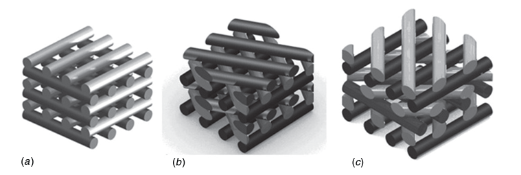
Header Image: Schematic representation of scaffolds characterized by three different lay-down patterns. (a) 0◦/90◦. (b) 0◦/60◦/120◦. (c) 0◦/45◦/90◦/135◦. [1]

The main objective of 3D bioprinting is to recreate human tissues with the same mechanical, structural and biological properties as the corresponding native tissue, in order to solve the problems associated with conventional transplantation techniques (donor site morbidity, organ shortage, etc.). For this purpose, different cell types combined with different biomaterials have been bioprinted according to specific models, but obtaining a 3D scaffold with the desired properties remains a challenge.

The advantage of 3D bioprinting over conventional scaffold fabrication techniques is the ability to control the 3D architecture of scaffolds through parameters such as pore size and geometry. Pore size and shape influence the resulting mechanical properties as well as cell behavior and tissue growth over time. Domingos et al. showed that for a lay down pattern of 0◦/90◦ (filament orientation between layers) with different pore sizes, poly(ε-caprolactone) scaffolds with smaller pores exhibit significantly higher stiffness under compressive conditions, which is an important property in applications such as bone tissue engineering. For different pore shapes with the following lay down pattern: 0◦/90◦, 0◦/60◦/120◦ and 0◦/45◦/90◦/135◦ (see figure) and the same porosity, the increasing number of angles between the filaments of the different layers leads to an increase in the deformability of the construct under compressive conditions.

Regarding the influence of architecture on cell behavior, viability and proliferation of human mesenchymal stem cells (hMSCs) were studied for 21 days and showed that larger pores with a lay down pattern of 0◦/90◦ improve viability and proliferation.

References

[1] Domingos, M., et al. “THE FIRST SYSTEMATIC ANALYSIS OF 3D RAPID PROTOTYPED POLY (e-CAPROLACTONE) SCAFFOLDS MANUFACTURED THROUGH BIOCELL PRINTING: EFFECT OF PORE SIZE AND GEOMETRY ON COMPRESSIVE MECHANICAL BEHAVIOR AND IN VITRO HMSC VIABILITY.” 1758-5082 5 (2013).

This article was written by Marie Moulin as part of an ongoing series of scientific communications written and curated by BioTrib’s Early Stage Researchers.

Marie is researching the Bioprinting of Bone and Cartilage at Uppsala University, Sweden.

Hip Arthroplasty Register

Total hip arthroplasty (THA) or total hip replacement is one of the most cost effective and reliable surgical operation. This operation consists in replacing the hip joint by prosthetic components allowing the patient suffering from hip pathology (ex: osteoarthritis) to restore painless motion and improve quality of life. For this surgical procedure, different models of implants are available (materials, shape, size and fixation methods) and surgeons decide depending on the age, pathology and medical history of the patient which implant characteristics would suit best. Joint implants are made to stay viable for the longest time possible in the body without revision surgery (second surgery related to an earlier inserted hip prosthesis). Revision surgery can occur after different complications like: repeated dislocation, infection or loosening of the implant and periprosthetic fracture [1].

In order to identify factor contributing to revision surgery and improve surgery procedure, national patient registries have been used in several countries. In 1979, Sweden was the first country to establish a national quality register collecting data on hip arthroplasty: the Swedish Hip Arthroplasty Register (SHAR). Nowadays a lot of countries possess regional and or national Hip Arthroplasty registers like Finland (1980), Norway (1989), Denmark (1995), Australia (1999), England, Wales, Northern Ireland and the Isle of Man (2002). The main objective is to centralize information within the country to follow the evolution of the number of total hip surgery, revision surgery as well as the prevalence in certain age group. Indeed, annual report are published to summarize data collected.

More importantly, registries are used to collect data on the patient, the surgical procedure and operation outcomes. The principal advantage is the possibility to investigate adverse outcomes of primary THA leading to revision surgery and improve surgical procedure. National registries play a major role in documenting the quality of THA to describe best practices and report outlier implants [2]. The 2019 Swedish Hip Arthroplasty Register report mention that “Never have so many hip arthroplasties been undertaken and never have so many research papers using data from the register been published during one operational year” [3].

References

[1] Varacallo M, Luo TD, Johanson NA. Total Hip Arthroplasty Techniques. 2022 Jul 4. In: StatPearls [Internet]. Treasure Island (FL): StatPearls Publishing; 2022 Jan.
[2] Varnum C, Pedersen AB, Rolfson O, Rogmark C, Furnes O, Hallan G, Mäkelä K, de Steiger R, Porter M, Overgaard S. Impact of hip arthroplasty registers on orthopaedic practice and perspectives for the future. EFORT Open Rev. 2019 Jun; 4(6):368-376. doi: 10.1302/2058-5241.4.180091.
[3] Kärrholm J, Rogmark C, Naucler E, Nåtman J, Vinblad J, Mohaddes M, Rolfson O. Swedish Hip Arthroplasty Register Annual report 2019. 2021 Feb. doi: 10.18158/H1BdmrOWu.

This article was written by Marie Moulin as part of an ongoing series of scientific communications written and curated by BioTrib’s Early Stage Researchers.

Marie is researching the Bioprinting of Bone and Cartilage at Uppsala University, Sweden.

Advances in non-invasive skin cancer imaging

Header Image:  Schematic in-vivo skin cancer imaging, with a synthetic bandwidth of 98 GHz using the designed ultra-wideband millimeter-wave imaging system.

Chances of early skin cancer treatment and intervention are enabled through tissue biopsy; prescribed by doctors when they suspect abnormal cell growth. The process itself requires doctors to harvest a sample of suspect tissue from individuals for external laboratory testing, leaving the patients with pain and wounds that take a long time to heal along with a period of uncertainty around cancer diagnosis whilst the sample is analysed.

However, a group of researchers from the Stevens Institute of Technology recently developed a technology to investigate abnormal tissues in-vivo and in real time by scanning a patient’s skin using millimetre-wave imaging technology, which is the same technology used in airport security scanners.

By examining 72 patients they were able to correctly differentiate benign and malignant lesions according to the way that the skin was reflecting light back. According to the research team, this happens due to the changes in the chemical composition of the cells. By utilizing an algorithm they are able to gather information and produce a 3D image in seconds even for the tiniest mole or imperfections indicative of cancer.

The device had a sensitivity and specificity of 97-98% and is comparable to even the greatest hospital-grade diagnostic instruments. Even though there are other devices available, those are not available in every clinic because of their size and cost. The technology that the team is developing is poised to integrate all the antennas and their circuits in a single chip making the device very small and low in cost.

References

Mirbeik, Amir, Robin Ashinoff, Tannya Jong, Allison Aued, and Negar Tavassolian. “Real-time high-resolution millimeter-wave imaging for in-vivo skin cancer diagnosis.” Scientific Reports 12, no. 1 (2022): 1-10.

This article was written by Edona Hyla as part of an ongoing series of scientific communications written and curated by BioTrib’s Early Stage Researchers.

Edona’s research is focussing on Understanding the Nature, Origin and Degradation of Implant Debris at the University of Leeds, UK

Is perfect research a myth?

One question pops into my mind, is it possible to conduct perfect research?

Then, I started reading some articles; one gripping article is presented here.

Whether it is possible to conduct perfect research or not is a controversial topic.

In 2005, Ioannidis wrote a paper titled “Why most published research findings are false” which is one of the most downloaded articles in PLOS Medicine. John Ioannidis employs mathematical concepts to demonstrate why published results are frequently incorrect. In simple terms, it’s about relying on significant p-values too often without considering the possibility of a false-positive result.

He listed six risk factors regarding the false results:

1. Small studies: “The smaller the studies conducted in a scientific field, the less likely the research findings are to be true”.

2. A small size of effect: “The smaller the effect sizes in a scientific field, the less likely the research findings are to be true”.

3. Testing many causal relationships simultaneously: “The greater the number and the lesser the selection of tested relationships in a scientific field, the less likely the research findings are to be true”.

4. Flexibility in study design: “The greater the flexibility in designs, definitions, outcomes, and analytical models in a scientific field, the less likely the research findings are to be true”.

5. Psychological bias: “The greater the financial and other interests and prejudices in a scientific field, the less likely the research findings are to be true”.

6. Hot research areas: “The hotter a scientific field (with more scientific teams involved), the less likely the research findings are to be true”.

When it comes to a query about whether we can improve the situation, he stated “it is impossible to know with 100% certainty what the truth is in any research question”. Rather we can work hard to improve the post-study probability. Finally, he suggested various countermeasures against the risk factor, concluding that research, by definition, is about making mistakes and striving to find a better approach. It is impossible to attain perfection. Many studies today indicate that he was correct.

What do you think?

This article was written by MM Raihan as part of an ongoing series of scientific communications written and curated by BioTrib’s Early Stage Researchers.

Raihan is researching In-situ Measurement of Nano-scale Wear Utilising Advanced Sensors at the University of Leeds, UK.