TriboUK BioTribology Posters

A strong attendance from Leeds biotribology researchers to the TriboUK 2022 conference hosted by Imperial College London.

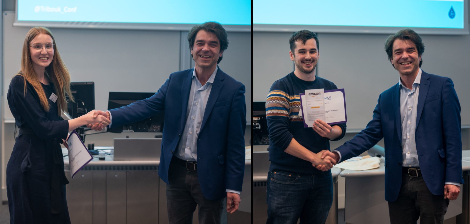
The University of Leeds walked away with both of the 1st prizes! Sarah Crossland won the top presentation prize for her talk on the measurement of foot plantar skin strain using digital image methods, and Rob Elkington won the top poster prize for his poster on polyelectrolyte functionalised PEEK for focal cartilage resurfacing.

Checkout the Leeds biotribology PhD student posters below from Sarah Crossland, Beril Yenigul, and Rob Elkington!

Sarah Crossland: Measurement of foot plantar skin strain using Digital Image Correlation methods for diabetic foot assessment

Beril Yenigul: Validation Rig Design and Results Design of a Facet Joint Replacement for Cervical Spine and Modification of a Hip Simulator for Simultaneous Friction Analysis and Validation of the Testing Method

Robert Elkington: Taking a PEEK at cartilage resurfacing

Osteoarthritis care (in)equity

Many of us believe that the extraordinary scientific advances that have led to the dramatic improvement of our health status are the prerogative of humanity. Unfortunately, the reality is quite different.

To give you some numbers, osteoarthritis affects 7% of the global population, more than 500 million people worldwide, with women disproportionately affected by the condition [1]. The number of persons affected worldwide increased by 48 percent from 1990 to 2019, and osteoarthritis was the 15th leading cause of years lived with disability (YLDs) [1]. Despite these numbers, many people with osteoarthritis still don’t get the first-line treatments (such as education and support for self-management, physical activity, exercise, and guidelines to maintain healthy body weight) and they are missing out on the care they need to live their lives to the fullest. For those particularly living in low- and middle-income countries (LMICs) this problem is exacerbated by the fact that both health-system and individual-level determinants influence access to care [2].

But let’s start in order. What is osteoarthritis? Osteoarthritis (OA) is the most common degenerative joint disease, a major cause of pain and disability, and a source of societal cost [3]. The disease compromises the structural and functional integrity of articular cartilage – the thick load-bearing tissue lining the ends of long bones – as well as the adjacent bone and other joint tissues [4]. The biological changes cause pain and limited mobility, which profoundly impacts a person’s everyday life, by restricting the ability to participate effectively in society, which can lead to social prejudice and exclusion from decision-making [2]. OA thereby leads to social, economic, and societal burdens, which are further compounded by the health challenges associated with increasing life expectancy and the prevalence of obesity in our global population [5].

While many of the barriers to providing OA treatment are universal, it is known that LMICs confront unique challenges and requirements. Although various difficulties have been raised, little is known about these obstacles and needs: disparity of care; expenses of providing and receiving treatment; and a lack of training for health workers [2].

The “Joint Effort Initiative” (JEI), an international consortium of doctors, researchers, and consumers led by the Osteoarthritis Research Society International (OARSI), was established with the goal of improving the global implementation of coordinated best-evidence osteoarthritis care. Driven by the huge impact of this chronic, complex condition, the JEI invited clinician-researchers from South Africa, Brazil, and Nepal to discuss their perspectives on challenges and opportunities in implementing the best-evidence care at the OARSI World Pre-Congress Workshop on April 28th 2021, to better understand some of the issues surrounding osteoarthritis care in LMICS [2]. There were five major themes that emerged when it came to overcoming obstacles to providing the best evidence-based osteoarthritis care [2]:

-Health inequities: refers to disparities in the ability of different groups of individuals to achieve their full health potential, which is commonly defined socially, economically, demographically, or geographically. In South Africa, Brazil, and Nepal, there are substantial links between health and wealth, with health disparities linked to high levels of poverty and aggravated by a lack of health insurance. For example, in rural areas of South Africa, access to healthcare is more limited. Furthermore, various political parties govern different provinces in South Africa, affecting the care accessible in each location and resulting in fragmentation of health services. In Brazil, it is estimated that over 18% of the population has poor access to healthcare, which rises to over 32% in rural areas. People from minority ethnic groups and those from lower socioeconomic strata have even less access. Many persons with OA in Nepal’s rural areas receive no care at all [2].

-Unaffordability of OA care: in both South Africa and Brazil, although the public health systems provide the majority of the OA care, there are significant wait times and insufficient support to help patients due to a lack of resources, so in Brazil for example people generally pay for their own OA therapy. In Nepal, since you must have health insurance to pay for OA treatment, many people simply do not have access to healthcare because it is too expensive [2].

-Lack of coordinated OA care: there is a dearth of specialized, coordinated osteoarthritis care, which is especially acute in rural locations [2].

-Unimportance of OA: OA is not considered an important disease and is often overlooked in low- and middle-income countries, where health budgets are restricted. For example, in South Africa, Brazil and Nepal, there is a scarcity of published OA research and no continuing national data collection for OA [2].

-Inexperienced: There is a general shortage of health professionals who provide OA therapy, and graduates are underprepared, particularly in rural areas [2].

But despite these major problems, efforts are being made for developing solutions and strategies to improve OA care in LMICs. In particular, these improvements are based on 3 major themes:

  • Upskill health workers via high-quality education and training: in South Africa, there are various training programs and the idea is to push these on a national basis in order to better integrate OA care into current health services. In Brazil, even if osteoarthritis is generally neglected, and in Nepal, they are also trying to educate and train health professionals through professional societies and universities to deliver best-evidence OA care [2].
  • Leverage national health priorities: since the treatments for osteoarthritis have not progressed as quickly as treatments for many other musculoskeletal and chronic noncommunicable disorders, the idea is to improve OA care by adapting successful practices and strategies from other areas of public health [2].
  • Use existing resources and advancements: to improve OA care, existing health breakthroughs and technologies could be expanded. For example, in these three countries, they are focusing on mobile health technology, so that they can reach a large part of the population quickly, even outside the urban area. This technology is already used for other diseases, so why not use it for osteoarthritis? In Nepal, furthermore, they are also trying to make more use of direct patient access to physiotherapists to manage pain [2].

In short, we cannot escape the bitter reality. Inequalities in osteoarthritis care between population groups exist in all countries, especially in low- and -middle-income countries. But fortunately, something is moving and despite many obstacles, as we can see from this article, there are also many opportunities and strategies for future implementations. The hope is that with these simple solutions, such as adopting strategies that have proven successful in other health conditions and providing education to health professionals and people with osteoarthritis [2], we can improve the care of osteoarthritis in the world and specifically in LMICs as soon as possible.

 

This article was written by Alessio Amicone as part of an ongoing series of scientific communications written and curated by BioTrib’s Early Stage Researchers.

Alessio is investigating the Elucidation of Friction-Induced Failure Mechanisms in Fibrous Collagenous Tissues at ETH Zürich, Switzerland.


References

[1] Hunter DJ, March L, Chew M. Osteoarthritis in 2020 and beyond: a Lancet Commission. Lancet. 2020 Nov 28;396(10264):1711-1712. Epub 2020 Nov 4.

[2] Eyles Jillian P., Sharma Saurab, Telles Rosa Weiss, Namane Mosedi, Hunter David J., Bowden Jocelyn L. Implementation of Best-Evidence Osteoarthritis Care: Perspectives on Challenges for, and Opportunities From, Low and Middle-Income Countries. Frontiers in Rehabilitation Sciences. 2022.

[3] Chen D, Shen J, Zhao W, et al. Osteoarthritis: toward a comprehensive understanding of pathological mechanism. Bone Res. 2017; 5:16044. Published 2017 Jan 17.

[4] Griffin, Timothy M.; Guilak, Farshid The Role of Mechanical Loading in the Onset and Progression of Osteoarthritis, Exercise and Sport Sciences Reviews: October 2005 – Volume 33 – Issue 4 – p 195-200

[5] Egloff C, Hügle T, Valderrabano V. Biomechanics and pathomechanisms of osteoarthritis. Swiss Med Wkly. 2012 Jul 19;142:w13583.



Thesis nailing: Unique Swedish tradition of PhD celebration.

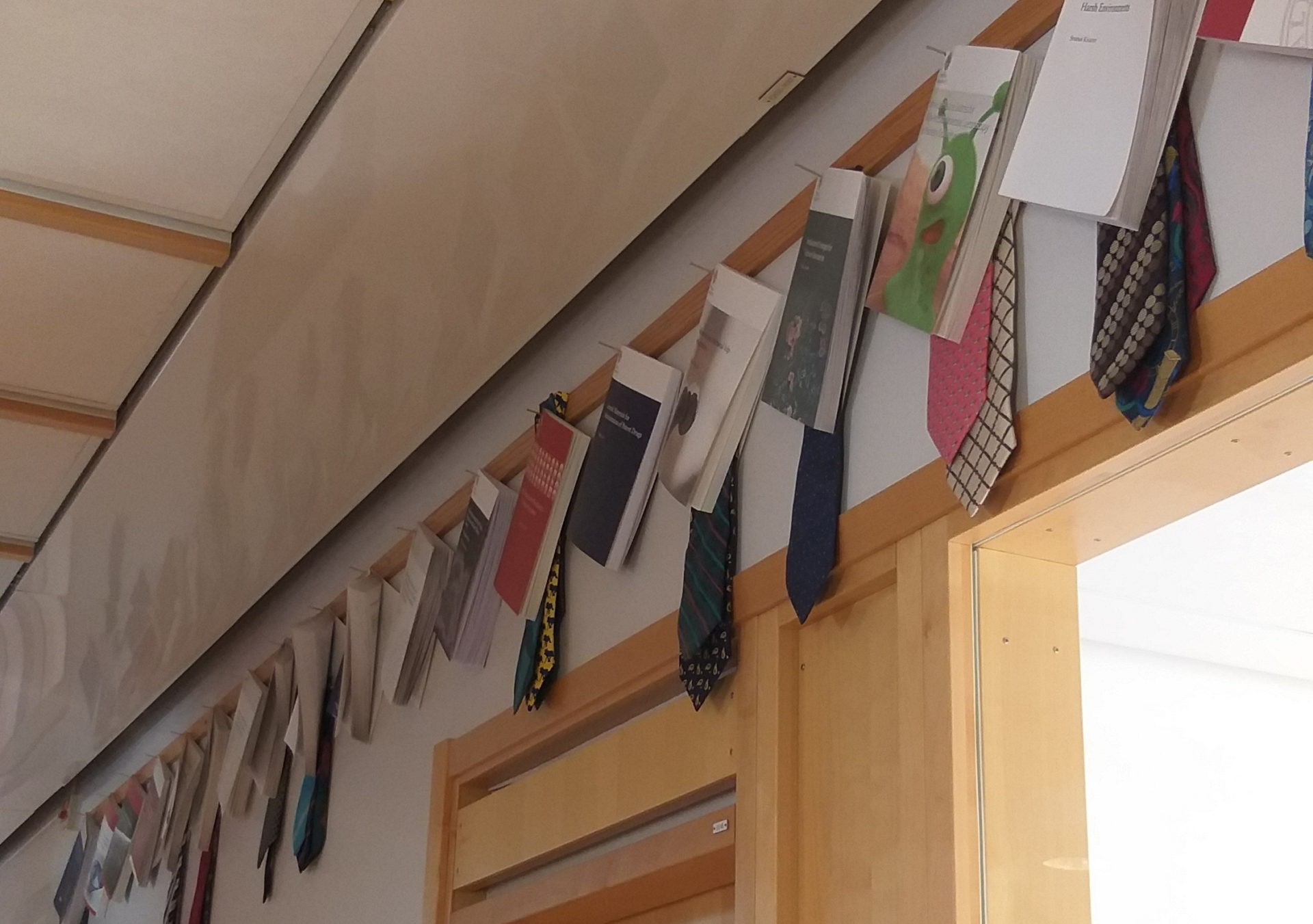
When I first entered Uppsala University’s engineering building, Ångströmlaboratoriet, I found one thing particularly interesting. Tens of books are nailed high up on the walls. Judging by the placement, I thought these books were obviously not for reading. Upon asking around, I learned that these books were printed doctorate theses that were nailed during the unique Swedish academic tradition called Spikning (or thesis nailing…literally!).

Several weeks before their thesis defense, the doctoral student announces the thesis nailing ritual and celebration. Usually, they will nail a physical copy of their thesis on the walls where their department resides. Of course, another copy will be nailed on an announcement board in the main hall of the building. A little ‘party’ comes after the nailing action, where the colleagues and supervisor can congratulate the doctoral student.

Figure 1. Nailed theses on a corridor of the applied materials science department.

It seems that this tradition has a long history. Inspiration for Spikning may have come from Martin Luther’s nailing of 95 theses on church doors in the 15th century [1]. Since various Christian denomination was already influential in Europe during that time, the idea does not seem too farfetched. If that is indeed true, then it just makes Spikning even more special. After all, I doubt we have many customs from the 15th century that is still a widespread practice now.

I find this unique Swedish academic tradition fascinating because it acts as both an announcement and a celebration. The journey to a doctoral degree is never easy. For many, I am sure it means years of commitment, effort, and perseverance. Thesis nailing offers doctoral students something extraordinary on their finish line: remembrance. The doctor may leave far from the university after the defense, but their thesis copy will stay long. In the corridor where my department resides, the thesis nailed on the walls dates as far back as the 1970s. Decades will pass, technologies will become obsolete, new knowledge will come, but something to remember the achievement will stay. I hope, by the end of BioTrib, I will be able to leave something that I can be proud of on a wall of Uppsala University.

Reference

[1]          “The (seeminlgy) strange custom of nailing PhD theses!,” Chalmeristbloggen, Mar. 09, 2015. https://chalmeristbloggen.wordpress.com/2015/03/09/the-seeminlgy-strange-custom-of-nailing-phd-theses/ (accessed Mar. 14, 2022).

This article was written by Vidhiaza Leviandhika as part of an ongoing series of scientific communications written and curated by BioTrib’s Early Stage Researchers.

Vidhiaza is researching the Development of Development of 3D-printed gradient alloys for joint implant component at Uppsala University, Sweden


Winner: Bioreactors and Growth Environments for Tissue Engineering

Mr. Manoj Rajankunte Mahadeshwara attended the course on ‘Bioreactors and Growth Environments for Tissue Engineering’ organized by Keele university. This interactive training course was spread across 2 days which targeted at postgraduate students (MSc and PhD) in industry and academia. Attendees were provided with a comprehensive understanding of the use of Bioreactors in Tissue Engineering. The course focused on bioreactors and growth environments for tissue engineering, covering bone, cartilage and connective tissues.

A bioreactor is a simulator that simulates the physiogical or pathological micro environment including physiological loading.

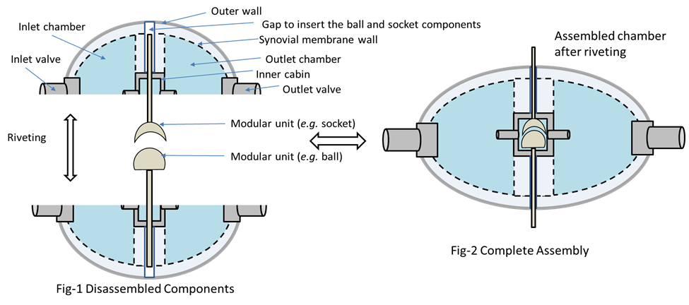
The winning BIOSYN reactor design for cartilage /osteoartis bioreactor.

He along with his team participated in the Bioreactor design challenge and has won the competition. His team designed the Bioreactor design for simulation of synovial joints – ‘BIOSYN’. This design was made to achieve three objectives which are:

  1. To investigate the cartilage-synovial fluid interface.
  2. To establish the effect of hydrostatic pressure of synovial fluid on cartilage/ tissue engineering construct
  3. To investigate how natural/artificial synovial fluid compositions affect cartilage (viability, differentiation status, etc.)


Manoj Rajankunte Mahadeshwara is currently a Tribos Erasmus joint Master’s student, pursuing a master’s in Tribology.

Manoj will be joining the University of Leeds in October 2022 for a PhD between the School of Dentistry and the Institute of Functional Surfaces (iFS) funded by a Bragg Centre for Materials Research PhD Scholarship.

Fika: Swedish tradition for ESR

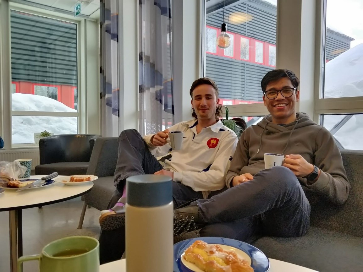
Fika (pronounced fee-ka) is a Swedish coffee break ritual. Swedes sit twice a day, about 10 a.m. in the morning and 3 p.m. in the afternoon. Fika is a Swedish word that roughly translates to “coffee and cake,” but it doesn’t quite capture the essence of the concept. “Fika” is an important component of socializing and maintaining a state of relaxation during the day and between jobs in Sweden, and you do not need to book it in your calendar.

In a nutshell, fika means ‘leaving your work behind and recharging your batteries.’

If you want to explore more about the essential Swedish ritual fika, there is a six-part web series (by Fabian Schmid) online. It is a way of life for some people, an institution for others, and a religion for others; it means different things to different individuals. But one thing is certain: in Sweden, it is a must for a healthy work-life balance.

People were advised to work from home when the covid-pandemic was at its peak, as they would be unable to randomly bump into someone or interact. Despite the pandemic, the Swedes continued to have fika with their coworkers, friends, and family (if in person, following the regulations in keeping the distance). They coined the term “virtual fika,” in which you sit in front of your camera with coffee, cake, and other refreshments and participate in fika.

Personally, I was astounded by the Swedish affection for fika. It not only gives you a respite from work, but it also improves your work efficiency by allowing you to refresh your thoughts for a few minutes. Furthermore, several studies have shown that it can significantly improve the bonding between colleagues in the workplace. Morley et al. [1] brilliantly discusses the fika and its role in today’s academic environment.

‘Fika allows my PhD students to communicate with me more freely and openly about project-related and, more significantly, personal matters. So that I may assist my students in any way I can,’ says Nazanin Emami, professor at Lulea University of Technology’s Division of Machine Element.

As an early-stage researcher, I can discuss my spontaneous project ideas with my colleagues and supervisor, discuss lab concerns, express my opinions on current events, cooperate with other colleagues, keep updates on other projects and and so on. This not only provides me an idea of what’s going on in the industry, but also about my colleague’s innovation and progress. Everything starts with fika and follows the formal protocol, whether it’s trying a new test approach or purchasing a new equipment for the lab. I find that having a moment of freshness helps me figure out where I am with my project. It can also enable your thought process to be redirected to a certain problem. Therefore, it helps to strengthen our relationship as colleagues.

References:

[1] Louise Morley, Petra Angervall, Caroline Berggren & Susanne Dodillet (2018) Re-purposing fika: rest, recreation or regulation in the neoliberalized Swedish University?, European Journal of Higher Education, 8:4, 400-414, DOI: 10.1080/21568235.2018.1458637


This article was written by Dilesh Raj Shrestha as part of an ongoing series of scientific communications written and curated by BioTrib’s Early Stage Researchers.

Dilesh is researching the Development of 3D-printable, self-lubricated polymer composites with improved wear resistance for total joint replacement at Luleå University of Technology, Sweden.


The Seven Secrets of Highly Successful Doctoral Students

I have recently attended a workshop called “The Seven Secrets of Highly Successful Doctoral Students”, held by Mr. Hugh Kearns. I would like to share these seven secrets with the research students. These seven secrets are as follows:

1. Care and maintenance of your supervisor

We should keep in mind that our supervisors are always busy and have many priorities along with our project. As it is our research, we need to become the driver. We should ask our supervisors to arrange meetings on a regular basis, even if we have done nothing! This way, we can use their valuable and practical tips and move our project forward. After each meeting, an agenda shall be prepared to indicate the following issues:

  • What I have done since last time
  • Questions/ issues
  • Feedback
  • What I will do next week
  • When is the next meeting?

2. Write and show as you go

Although it seems time-consuming, it is required that the research students regularly write since writing is a creative process clarifying thinking as well as developing ideas. We usually like to write when we feel we are ready, but we may never feel ready; therefore, we should write early, preferably in the morning, on a regular basis. Furthermore, we should get feedback from our supervisors and peers as writing is not improved by itself.

3. Be realistic

4. Say no to distractions

Social media is the number one distraction for wasting our time and for not doing work.

5. It’s a job

Although we should work for a certain amount of time daily, we have holidays as well. Thus, if we specify at what times we should focus on our work and when we do not have to work, we will get more done.

6. Get help

7. You can do it

One of the primary things that can help us significantly is perseverance, and the role of hardworking in making better progress is much more critical than intelligence.


This article was written by Mahdieh Mosayebias part of an ongoing series of scientific communications written and curated by BioTrib’s Early Stage Researchers.

Mahdieh is researching the Design of Self Lubricating Prothesis at ETH Zurich, Switzerland.

Post assessment of Total Hip Replacement Patient: An integrated AE measurement Technique

For many years, joint replacement of damaged hips has been a standard treatment in orthopaedic surgery. There is currently no mechanism for early detection of implant failure following surgery. Early detection of total hip replacement (THR) failure could lead to more proactive surgical intervention and better patient outcomes. In this instance, the Acoustic Emission (AE) measuring approach may be a good fit as a diagnostic indication for joint health, implant failure modes, and gait analysis.

The previous study’s AE monitoring technique did not collect patient motion data, making it hard to make accurate comparisons between AE events and implant motions, or to determine whether specific AEs are caused by specific implant articulation angles, loads, or angular velocities. FitzPatrick et al. (2022) established a concurrent technique of AE monitoring to combine lower-limb motion and AE data to enable temporal interpretation of acoustic information for gait analysis to study this issue.

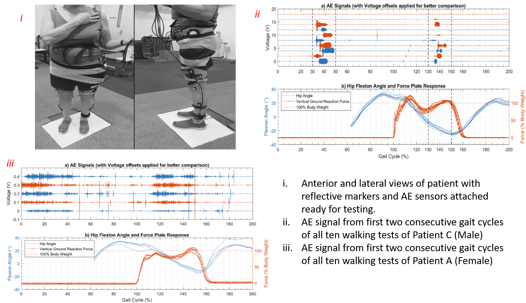
Three patients (two males and one female) between the ages of 50 and 70 were taken for a combined AE and gait analysis. They underwent a ceramic-on-ceramic implant bearing hip replacement. Four passive ultrasonic receivers set in a flexible array on the patient’s skin surface from the iliac crest to the upper femur were used to identify AEs. As the patient walked across the room in a straight line at a self-selected speed over a force plate, AE data were recorded. A motion analysis system with six infrared tracking cameras recorded the patient’s limb motions at the same time.

Their findings revealed that AEs are significantly linked to the stance phase of walking, when implant loads are high and the hip joint’s angular velocity is high. The key observation from the male patient was that all of the recorded squeaks happened between 30% and 50% of their individual gait cycle, which pertains to terminal stance, across all walking tests. Interestingly, the situation was significantly different for the female patient; total voltage magnitudes were lowered, and AEs of significant magnitude occurred consistently during the stance phase.

Based on current findings, the exact mechanism that causes implant squeaking is unknown. As a result, ongoing research aims to collect combined AE and gait data from more hip replacement patients in order to evaluate if the findings apply to a larger group of patients and to get greater insight into quantitative relationships between AE activity and hip joint dynamics.

Source: FitzPatrick, A. J., et al. “Synchronized acoustic emission and gait analysis of total hip replacement patients.” Biomedical Signal Processing and Control 74 (2022): 103488.


This article was written by MM Raihan as part of an ongoing series of scientific communications written and curated by BioTrib’s Early Stage Researchers.

Raihan is researching In-situ Measurement of Nano-scale Wear Utilising Advanced Sensors at University of Leeds, UK.

International Womens Day – BioTrib

For International Women’s Day 2022 the women in BioTrib have put together a series of 6 interviews and articles covering:

– Why engineering as a profession?
– Women of Impact: Empowered women, empower women. 
– What did you expect your experience of engineering to be like, and how does that compare to reality?
– What skill(s) in particular have helped you during your career?
– What advice would you give to your younger self about entering STEM?
– Do you think that the proportion of women in your field has changed over the course of your career?

Thanks for editing and contributions from Judith SchneiderCecilia PerssonEdona HylaIsobel ReesBeril Saadet YenigülAfrina Khan PiyaDr Lisa-Dionne MorrisFjolla SylajIsobel Pollock-Hulf OBE and Charlotte Merrell

Check it out below!

This project has received funding from the European Union’s Horizon 2020 research and innovation programme under the Marie Skłodowska-Curie grant agreement No 956004 🇪🇺

Ukraine: In the face of adversity the international science community brigades

Ever since Russian invasion of Ukraine we have seen a rise in researchers & scientists coming out in solidarity with Ukrainian students and researchers by sharing opportunities in their universities and labs for them.

One of the initial such opportunities posted on Linkedin by Prof. Yiannis Pontikes reached nearly 150,000 people and is still gaining traction. This led to other scientists posting similar opportunities for the Ukrainian students and researchers leading to the launch of global hashtag #ScienceForUkraine that has been trending on Twitter and Linkedin where scientists throughout the world are sharing opportunities for Ukrainian students to continue their research.

A further online spreadsheet (link: Labs supporting Ukrainian Students) by Andrew Kern @pastramimachine has been created to help Ukrainian scientists locate the professors and funding departments throughout the world at all career levels. There is an interactive website Science for Ukraine to serve the same purpose. These lists are continuously updated and any academic interested in coming forward to help Ukrainian students and researchers can put their details. The list contains more than 500 opportunities at the moment and it is growing quickly.

Alliance of Science Organisations in Germany (DFG) have in their press release announced support for students and researchers from Ukraine under wide-ranging assistance programs being announced or to be announced. Polish academy of Sciences have also launched a website to help Ukrainian students and researchers with funding as well as further support to find a supervisor in their area of research.

Many other EU countries have also launched similar initiatives to support Ukrainians.

We are stronger together, in the face of this adversity the global scientific community has showed that while there may be war, the only way forward for future of science is through support and collaboration at a global scale.


This article was written by Sallar Ali Qazi as part of an ongoing series of scientific communications written and curated by BioTrib’s Early Stage Researchers.

Sallar Ali Qazi is researching Mechanical and Tribo-Chemical Wear Modelling of Artificial Joint Prostheses at Imperial College London, UK

LGBT History month: Alan Turing History and a Call to action

Alan Turing was certainly one man ahead of his time. He was fascinated by mathematics and logic and laid the foundations for what would become modern computer science. He helped the United Kingdom in the war efforts to break the Enigma, a dreaded cryptography machine used by Nazi Germany for communications. Later, he would also come up with concepts that just now are being explored such as artificial intelligence and mathematical biology (BBC Horizon).

Turing was homosexual, for this sole reason, he was arrested in 1952 for indecency. He was chemically castrated and had developed a depression that might have caused his suicide (Doan, 2017). Doan (2017) reflects on his tragic fate as a classic example of how society’s prejudice robbed him of a dignified and fulfilling life.

We might have progressed in tolerance and respect of LGBTQ+ community in most countries; however, LGBTQ+ scientists are still more likely to suffer discrimination in the workplace. This culminates in a higher likelihood of depression, stress at work, insomnia, and other health issues (ELSE, 2021). Specially LGBTQ+ ethnical minorities and women are subject to the effects of prejudice (ELSE, 2021).

Let us not forget this tragic example and keep fighting to promote a more egalitarian culture in honor of Turing’s and so many lives wasted to intolerance.  As scientists, we can advocate for more inclusive and respectful workplaces and societies for everyone, regardless of gender expression and sexual orientation.


This article was written by André Plath as part of a series on LGBTQIA+ History Month. curated by BioTrib’s Early Stage Researchers.

André is one of BioTrib’s Early Stage Researcher‘s who is investigating Boundary Lubrication of Fibrous Scaffolds at ETH Zürich, Switzerland.


Header Image: “Alan Turing” by avaragado is licensed under Creative Commons. Image source: https://wordpress.org/openverse/image/a6514d5d-a295-4a60-8023-9a3e89f81c1b

References

Alan Turing BBC Horizon. Available at: <https://www.youtube.com/watch?v=Z-sTs2o0VuY>

Doan, L. (2017). Queer History / Queer Memory. In GLQ: A Journal of Lesbian and Gay Studies (Vol. 23, Issue 1, pp. 113–136). Duke University Press. https://doi.org/10.1215/10642684-3672321

Else, H. (2021). The largest-ever survey exposes career obstacles for LGBTQ scientists. In Nature. Springer Science and Business Media LLC. https://doi.org/10.1038/d41586-021-00221-w


Polymer Brushes and Lubrication: Nature Inspires New Biomaterial Advances

PRG4 or lubricin is a protein with a bottle-brush shape that can be perfectly mimicked by polymer brush grafting to biomaterial surfaces. This imparts to biomaterials’ surfaces super lubricous properties and a coefficient of friction (µ) lower than 0.01.  In addition, polymer brushes grafted to material surfaces may impart tunable hydrophilicity, self-cleaning, catalysis, controlled cell, and bacteria adhesion [1]. They can be applied for response actuation and drug delivery. Lubrication polymer brushes can be charged (positive and negative charges), amphiphilic, or act via steric hindrances[1]. The end properties can be controlled by molecular weight, grafting density, and radius of gyration.

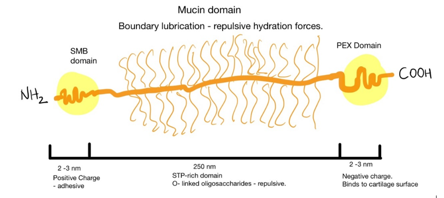
One of the mechanisms of lubrication is brush hydration. The thick water film would prevent the probe from contacting the surface[1]. In nature, this role is played by hyaluronic acid and other sugar molecules in articular cartilage, for example. The sugar molecules are conjugated to lubricin forming a mucinous domain. The protein is anchored by a somatomedin-B (SMB) domain to hyaluronic acid from the extracellular matrix of cartilage cells (chondrocytes) [2,3]. The glycosylated domains can trap water and establish electrostatic repulsion promoting lubricity of the tissue [4,5]

Figure 1: Lubricin domains

Several strategies have been developed to mimic this biochemical environment. Poly(l-lysine) (PLL) brushes were grafted onto poly(ethylene glycol) (PEG) surfaces to obtain good lubricity and biocompatibility[6]. Morgese et al. grafted to poly(glutamic acid) different polyoxazolines. These polymers are known for passivating surfaces and promoting now-fouling without eliciting immune responses. In samples with hydroxybutyrate (HBA), she obtained a biomimetic material that could bind to degraded cartilage and regenerate tissue[7]. This implies interesting solutions for people with early-onset arthritis.

To conclude, bottle-brush materials might be the future of cartilage tissue engineering, but more studies need to be conducted to show the in vivo feasibility of the concepts. We expect these new materials to influence scaffolds/gels potentially entering the market in the next decades.

REFERENCES

[1]          S. Ma, X. Zhang, B. Yu, F. Zhou, Brushing up functional materials, NPG Asia Mater. 11 (2019) 24. https://doi.org/10.1038/s41427-019-0121-2.

[2]          Y. Lee, J. Choi, N.S. Hwang, Regulation of lubricin for functional cartilage tissue regeneration: a review, Biomater Res. 22 (2018) 9. https://doi.org/10.1186/s40824-018-0118-x.

[3]          I. Bayer, Advances in Tribology of Lubricin and Lubricin-Like Synthetic Polymer Nanostructures, Lubricants. 6 (2018) 30. https://doi.org/10.3390/lubricants6020030.

[4]          S.M.T. Chan, C.P. Neu, G. DuRaine, K. Komvopoulos, A.H. Reddi, Atomic force microscope investigation of the boundary-lubricant layer in articular cartilage, Osteoarthritis and Cartilage. 18 (2010) 956–963. https://doi.org/10.1016/j.joca.2010.03.012.

[5]          I.M. Schwarz, B.A. Hills, Surface-active phospholipid as the lubricating component of lubricin, Rheumatology. 37 (1998) 21–26. https://doi.org/10.1093/rheumatology/37.1.21.

[6]          S. Lee, M. Müller, M. Ratoi-Salagean, J. Vörös, S. Pasche, S.M. De Paul, H.A. Spikes, M. Textor, N.D. Spencer, Boundary Lubrication of Oxide Surfaces by Poly(L-lysine)-g-poly(ethylene glycol) (PLL-g-PEG) in Aqueous Media, Tribology Letters. 15 (2003) 231–239. https://doi.org/10.1023/A:1024861119372.

[7]          G. Morgese, E. Cavalli, J.-G. Rosenboom, M. Zenobi-Wong, E.M. Benetti, Cyclic Polymer Grafts That Lubricate and Protect Damaged Cartilage, Angew. Chem. 130 (2018) 1637–1642. https://doi.org/10.1002/ange.201712534.


This article was written by André Plath as part of an ongoing series of scientific communications written and curated by BioTrib’s Early Stage Researchers.

André is researching Boundary Lubrication of Fibrous Scaffolds at ETH Zürich, Switzerland.

LGBTQ+ Researcher Visbility: 500 queer scientists

500 queer scientists (Actually 1,625+ queer scientists) is a visibility campaign for LGBTQ+ and allied people working in STEM and STEM supporting roles. It is a database of self-submitted biographies and stories intended to boost recognition and awareness of STEM scientists. This is with the view of helping isolated members of the queer community realise they are not alone and perhaps even create opportunities and connect communities in academic or professional institutions!

Visibility for LGBTQ+ STEM workers is critical for cultivating wellbeing in professional and academic environments. Many members of the LGBTQ+ community have reported incidents of harassment and discrimination in STEM environments,

It is estimated LGBT people are approximately 20% less represented in STEM fields than expected [Cech, 2017]. With nearly 28% of LGBT and 50% of trans staff at least once considering leaving the workplace due to a climate of discrimination [RSC, IOP 2019].

Further statistics and information is available on the 500 QS resource page.

If you are an LGBTQ+ person or ally in the STEM community, you can help grow 500 QS by submitting your own biography!


References

RSC, IOP 2019: https://www.rsc.org/globalassets/04-campaigning-outreach/campaigning/lgbt-report/lgbt-report_web.pdf

Cech, 2017: https://doi.org/10.3390/socsci6010012 


This article was written by Rob Elkington, the BioTrib website manager as part of a series of blog posts for LGBTQ+ history month.

 

BioTrib Silk Road – Embrace our opportunity of international research collaboration

Reaching back over 2,000 years, an ancient network of trade routes called the ‘Silk Road’ connected the East and the West. Precious goods, splendid cultures and religions travelling along thousands of miles, stroke, exchanged and merged. The term ‘Silk Road’ was first used by German geographer Ferdinand von Richthofen in 1877, as silk is one of the favourite goods traded from China to Europe, also as a metaphor for the ideas travelled from different civilizations.2

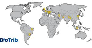
Amazed by this picture (marks are the origins of all ESRs who joined BioTrib this year) and the idea of ‘BioTrib Silk Road’ presented by Prof Richard during our ESRs meeting, I started to think about the importance of international research collaboration in the modern world.

BioTrib Silk Road – Image by Prof Richard M Hall, recreated by Esperanza Shi


“Ideas transcend borders, no country controls the marketplace of ideas.”

— Alejandro Adem 3

Indeed, there isn’t a researcher who knows everything in the world, nor a university owns all of the state-of-the-art equipment and facilities. We have to collaborate, and we love to collaborate. When people from diverse backgrounds meet, idea sparks. When institutions collaborate, science thrives. While in BioTrib, deep international connections have formed between universities and industries from the UK, Sweden, Switzerland, Germany, China and Australia; researchers are not only from different academic backgrounds but also diverse cultural backgrounds. The diversity and inclusiveness are the treasures of BioTrib and I can’t wait to see our footprints of contribution to academic research on this ‘BioTrib Silk Road’.

Header Image: Marco Polo Geography and Map Division/Library of Congress, Washington, D.C. (gct00215-ca000005) 1


References

(1) Marco Polo on the Silk Road https://www.britannica.com/topic/Silk-Road-trade-route

(2) The Silk Road https://www.nationalgeographic.org/encyclopedia/silk-road/

(3) The benefits and challenges of international research collaboration https://www.universityaffairs.ca/features/feature-article/the-benefits-and-challenges-of-international-research-collaboration/


This post was written by Esperanza Shi as part of an ongoing series of scientific communications written and curated by BioTrib’s Early Stage Researchers.
Esperanza is researching the Optimisation of Scanning Strategies for 3D Printed Artificial Joints at Imperial College London, UK.


Advances in Additive Manufacturing: 3D-printed microneedles

The ongoing 4th Industrial Revolution has shifted the traditional paradigm of producing medical devices. Additive Manufacturing (AM), a mould-less technology commonly referred to as 3D printing, plays an essential role in the shift taking place in this field.

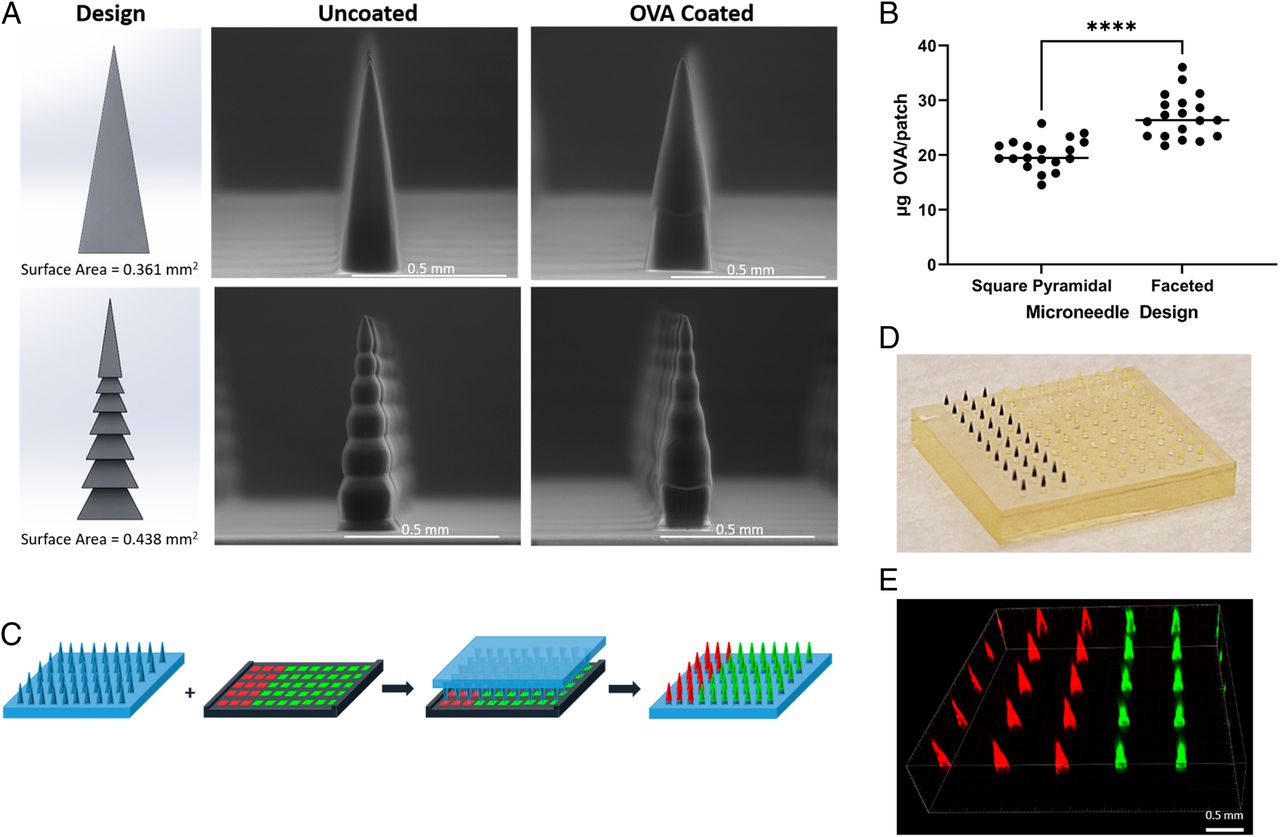
Because of the high degree of geometrical freedom that can be achieved, AM is being used to conceive polymeric microneedles (MNs) with tailored design. For instance, Caudill and co-workers (2021) studied the benefits of microneedle vaccination over the traditional subcutaneous one. An AM process that relies on resin photopolymerization (i.e., continuous liquid interface production) was used to fabricate the MNs in two different shapes: square pyramidal and faceted (cf. image given in this post).

Cargo loading was performed via surface coating and assessed for the different MN designs. Whilst surface area increased 21.3%, cargo loading augmented 36% from square pyramidal to faceted with horizontal grooves, which pinpoints the importance of geometry design to loading biologics on MNs. Furthermore, transdermal delivery through MN vaccination was more effective in triggering primary antigen-specific IgG as well response duration when compared to subcutaneously or intradermally delivering paths.

Caudill et al. (2021) findings represent a major step towards a simpler, effective, and pain-free vaccination process that can potentially increase global vaccination. Furthermore, this self-administered vaccination path may aid in prompt responses during epidemic and pandemic scenarios. In that sense, AM has proved to be a feasible manufacturing route for improving drug delivery systems via tailored shapes and geometries.

Read more of this fascinating paper here: Transdermal vaccination via 3D-printed microneedles induces potent humoral and cellular immunity

This post was written by Pedro Luiz Lima dos Santos as part of an ongoing series of scientific communications written and curated by BioTrib’s Early Stage Researchers.
Pedro is researching the Functional Biotribology of the Surface Engineering of 3D Printed Components at the University of Leeds, UK.

BioTrib collaborators; University of Leeds and Imperial College, together with ETH Zurich and Uppsala University as project partners are awarded a programme grant for the treatment for spinal metastases

A £7 million research project has been launched to develop a new imaging and keyhole surgery approach to the treatment for secondary bone tumours of the spine. 

Known as metastatic bone disease, the tumours spread from a primary cancer located elsewhere in the body. The condition is particularly associated with breast cancer.  

The bone tumours cause vertebrae to weaken and eventually fracture, leaving people in severe pain, immobility and requiring surgery. In some cases, the fracture may damage the spinal cord and cause paralysis. For these patients, however, quality of life is a key issue and complex surgery may be inappropriate. 

A research collaboration between the University of Leeds, Imperial College London and UCL has received funding to develop an alternative approach based on developing new imaging and modelling techniques that will enable clinicians to predict which patients are at a high-risk of a vertebra fracturing.  

They would then be fitted – using minimally invasive surgery – with a tailor-made implant to strengthen the spine and prevent the fracture. 

The project – Oncological Engineering: A new concept in the treatment of bone metastases – has attracted £7 million in research funding, including £5.6 million grant from the Engineering and Physical Sciences Research Council (EPSRC), part of UK Research and Innovation, the Government-funded body set up to support research and innovation. 

“The problem facing doctors is they have no way of knowing which of the spinal vertebrae is going to collapse. But when that happens, patients may require major surgery which involves a lengthy period of rehabilitation. 

Our approach is to intervene by developing new techniques and equipment that will prevent spinal fractures, crucially helping to maintain a patient’s quality of life at a time when they may be terminally ill. ”

Professor Richard Hall

BioTrib Coordinator and expert in medical engineering at the University of Leeds who is leading the new research collaboration

According to Cancer Research UK, 150 people every day are diagnosed with breast cancer. Although more than 76% of people with the disease survive for more than ten years, some patients do develop stage 4 cancers, of which it is estimated about 50% to 60% get bone tumours.  

In stage 4 cancers, the disease has spread to other organs. 

Within five years, the research team hope to have developed new techniques and materials that will revolutionise the treatment of bone metastases.  

The approach is based on personalised medicine, assessing an individual’s risk that the spine has weakened so much that a vertebra will fracture. In those cases where surgeons intervene to strengthen the spine, the implant will be tailor-made.  

“Through improvements in imaging and modelling and a personalised approach, this project has the potential to revolutionise the treatment of secondary bone tumours. 

It demonstrates the importance of fundamental research and engineering solutions in developing new treatments that will have a profound impact on peoples’ lives.” 

Dr Kedar Pandya

Director for Cross-Council Programmes at the Engineering and Physical Sciences Research Council

Predicting the risk of a vertebrae fracturing 

Researchers will develop news approaches to patient imaging and computer modelling, enabling them to track tumour development in the spine over time and how it might be weakening individual vertebrae. The information would be compared with the loading on the spine, enabling clinicians to predict which of the vertebrae is at risk of fracturing. 

“This funding will enable us to significantly expand our work combining computational modelling with cutting-edge imaging to better understand how cancers grow and interact with surrounding tissues.  

We are excited to use these multidisciplinary frameworks to understand vertebra fracture risk and ultimately help to improve quality of life for cancer patients.” 

Professor Rebecca Shipley

Department of Mechanical Engineering at UCL and one of the co-investigators

Implant made of advanced materials 

Those vertebra at a high-risk of collapse would be supported by an implant inserted into the spine using minimally invasive techniques.  

The implant would be made from what is called a metamaterial, a material that has uncommon properties that can be fine-tuned to the needs of the patient, for example the material could harden when under stress. 

Metamaterials are currently used in the aerospace industry but with advances in 3-D computer printing, the research team believe they could be adapted to provide tailor-made structural integrity to vertebrae at high risk of fracturing. 

The advanced manufacturing group from the Dyson School of Design Engineering at Imperial College, London, will be developing a novel 3D printer capable of fabricating the intricate implant designs. Their machine will utilise smart optical systems to print photopolymers at extremely fine resolution. 

“This project allows us to expand our expertise in the analysis, optimisation and 3D printing of structural metamaterials. By working as part of the multidisciplinary team we aim to apply the new approaches and knowledge to improve the quality of life of late-stage cancer suffers.  

We will also be able to apply some of these new approaches back into the aerospace and mechanical engineering sectors where advanced meta-materials have a wide range of potential applications.”

Dr Rob Hewson

BioTrib Lead Scientist and Co-investigator of new research collaboration at Imperial College

By using minimally-invasive techniques to implant the material, the recovery period for patients will be days – rather than weeks or months with the surgery that is required if one of the spinal bones fractures. 

The NHS long-term plan for cancer treatment had called on researchers to develop new interventions that would improve the quality of life of patients living with advanced cancers. 

It is hoped the new techniques will be applied to other areas of the healthcare sector.