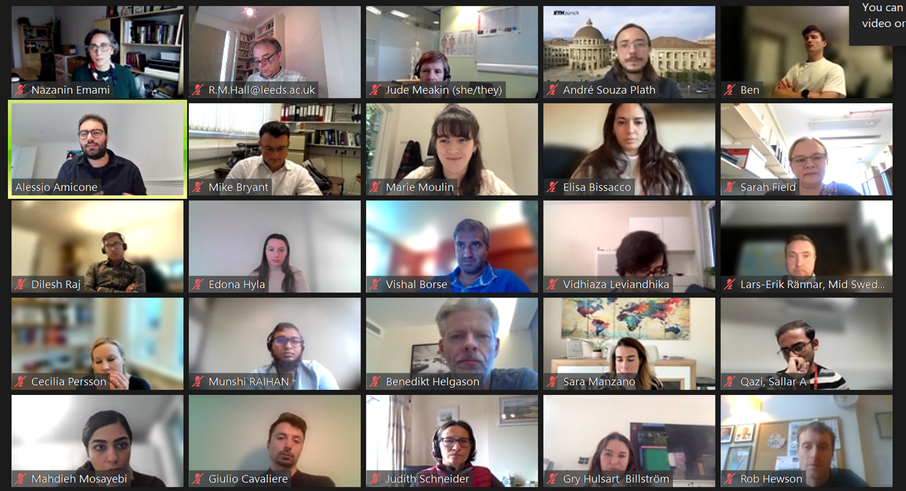
Great recent External Expert Advisory Board meeting for BioTrib!
Early Stage Researchers provided an overview of the progression in each of the work packages and their own career development.
Erudite conversations, succinct presentations and insightful recommendations from the board. Special thanks to the external advisory members including the Chair, Dr Vishal Borse, Jude Meakin, Sara Manzano, Heather Yates and Lars-Erik Rannar.
Header Image: A sample of the BioTrib personnel present at the EEAB.
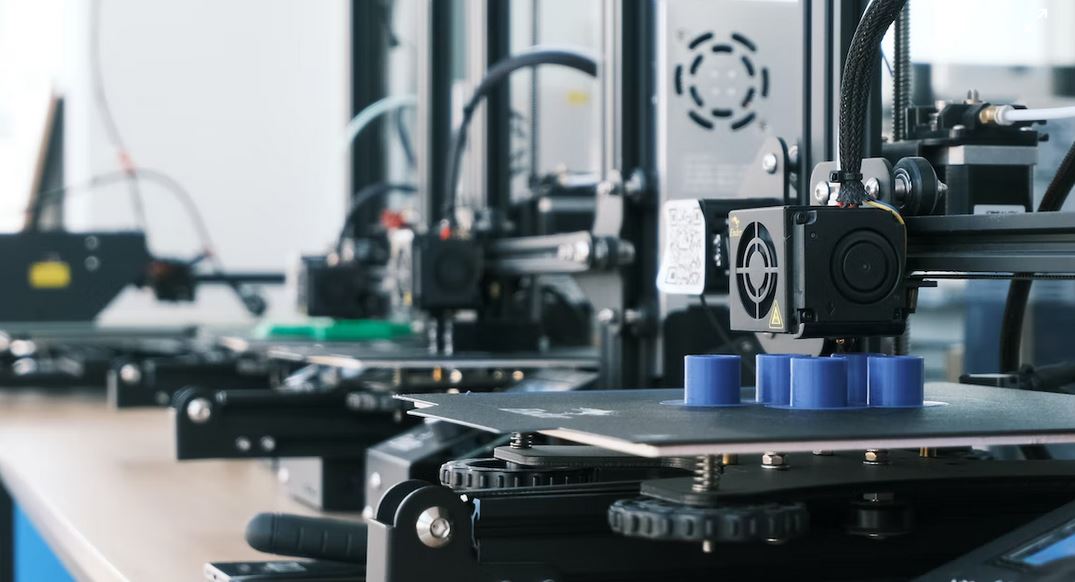
Additive Manufacturing (AM) is the process of creating an object by building it one layer at a time. It is a sustainable production method that eliminates the need for excess material and unnecessary waste. It can be used to fabricate complex shapes without tooling. The cost of manufacturing with AM is often not cheaper than conventional processing, particularly if the product is designed for mass production. AM processes include VAT photopolymerization, material jetting, material extrusion, binder jetting, powder bed fusion, sheet lamination, and directed energy deposition. These AM processes are briefly explained as follows:
VAT photopolymerization: liquid photopolymer in a vat is cured by light, and the material form is liquid.
Material jetting: droplets of material are selectively deposited, and the material form is liquid.
Material extrusion: material is dispensed through a nozzle in layers, and the material forms are filament, pellets, and paste.
Binder jetting: a liquid bonding agent is selectively deposited, and the material form is powder.
Powder bed fusion: thermal energy fuses regions of a powder bed, and the material form is powder.
Sheet lamination: sheets of material are bonded to form an object, and the material form is sheets.
Directed energy deposition: focused thermal energy fuses materials as deposited, and the material forms are powder and wire.
The medical applications of AM are classified as follows:
Medical models: Medical models are based on patient anatomy, and they can be used for preoperative as well as postoperative planning. They are widely used in the craniomaxillofacial area, also for limbs, the spine, and the pelvis. Powder bed fusion, material extrusion, and binder jetting are three AM processes that are usually utilized for medical models.
Implants: Implants are manufactured directly or indirectly by AM to replace damaged or missing tissue. AM is a favorable choice for manufacturing personalized implants. Most of the implants are constructed from metals using the powder bed fusion process. In some cases, polymers and ceramics are utilized to fabricate the implants.
Tools, instruments, and parts for medical devices: Tools, instruments, and parts for medical devices improve clinical operations. Patient-specific dimensions and shapes might be used. Surgical instruments as well as orthodontic appliances can be considered in this classification. The VAT photopolymerization process is often employed to manufacture tools, instruments, and parts for medical devices.
Medical aids, supportive guides, splints, and prostheses: Parts created with AM are external to the body and can be customized. Long-term and postoperative supports, motion guides, fixators, external prostheses, prosthesis sockets, and personalized splints can be taken as examples of this category.
Biomanufacturing: A combination of AM and tissue engineering is considered Biomanufacturing which mostly uses polymers, ceramics, and composites. Porous structures are also utilized that are favorable to attracting cells and cell growth.
Reference:
[1] Salmi, Mika. “Additive manufacturing processes in medical applications.” Materials 14.1 (2021): 191.
This article was written by Mahdieh Mosayebias part of an ongoing series of scientific communications written and curated by BioTrib’s Early Stage Researchers.
Mahdieh is researching the Design of Self Lubricating Prothesis at ETH Zurich, Switzerland.
Sometimes unexpected events occur at unexpected times, providing inspiration for medicine. In this instance, it pertains to ophthalmology, more specifically intraocular lenses, and it occurred during World War II. Intraocular Lenses (IOLs) are optical lenses that can correct refractive defects by being implanted in the eye. IOL-based cataract surgeries are currently one of the most popular and safest surgical procedures performed globally. (Yu, 2018)
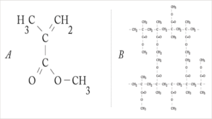
The Second World War did, in fact, mark the genuine commencement of the IOL. Harold Ridley was a civilian ophthalmologist, who were operating on Royal Air Force pilots who suffered eye injuries, engaged in aerial combat over the South of England. On August 15, 1940, he underwent 19 surgeries on a pilot, whose Perspex canopy had shattered, sending numerous splinters of poly(methyl methacrylate) (PMMA, Figure 1) into his eyes.
Figure 1. (a) MMA (methyl methacrylate) forms the basis for acrylic IOLs. (b) (PMMA) is a transparent thermoplastic.
He discovered that the PMMA splinters, unlike glass splinters, remained inert in the patient’s eye and that the immune system had not responded to them during the course of the treatment. Ridley came to the realisation that PMMA could be utilised to create artificial lenses that could be inserted into the eye to replace the natural lenses that had been removed during cataract surgery. (Kretz, 2014)
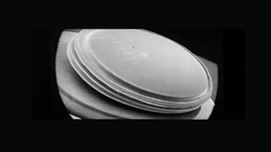
He then, together with the Rayner Optical Company, produced the first IOL (Figure 2) adopting ICI’s Transpex I, a high quality version of PMMA. On November 29, 1949, at St Thomas’ Hospital, London, Ridley performed surgery inserting the first lens into a 42-year-old woman after an extracapsular cataract excision. (Scholtz, 2006)
Figure 2. Scanning electron micrograph of a Ridley intraocular lens made from PMMA
Today, millions of people’s lives have been made better thanks to Ridley’s brilliance. IOLs are now widely used for refractive outcomes improvement throughout clear lens exchange, not just during cataract surgery. The quality of vision following cataract surgery has never been greater in the developed world thanks to the incremental advancements in IOL design.
Take-Home Message: always keep an open mind, you never know where the next important medical discovery might come from!
References
Yu, N. F. (2018). State of the art of intraocular lens manufacturing. The International Journal of Advanced Manufacturing Technology, 1103–1130.
This article was written by Elisa Bissacco as part of an ongoing series of scientific communications written and curated by BioTrib’s Early Stage Researchers.
Would you accept an organ transplant from an animal donor? Nature’s recent paper “ Will pigs solve the organ crisis?” [1] sparked this question in my brain. On one hand, animal-grown organs might significantly reduce transplantation waiting times. On the other hand, practical and ethical concerns arise from genetically modified animals, cruelty, organ rejection, and infections.
According to Organ donor, 17 people die on the organ transplant waiting list each day [2]. In the US, more than 100.000 people are waiting for a transplant. Kidney, liver, and heart are the most transplanted organs. Approximately 35.5 thousand transplants were performed between the months of January to October 22 [3]. That means only approximately one in 3 people will receive a much-needed transplant. These statistics do not reflect the global reality – even in some countries, regional differences may influence the likelihood of a patient getting a needed new organ.
Could animal-grown organs be the solution to long-transplant lines?
Genetic modifications are allowing size-compatible organs, lack of immune rejection, and thus increased lifespans [1]. However, the biggest limitation lies in the increased risk of virus spillover from animals to humans and the presence of endogenous retroviruses that might be harmful to the patient [1]. Polemic issues with human testing on brain-dead patients were also pointed out by the paper [1]. From the animals’ perspective, it is also important to assure that genetic modification does not impart suffering. They must also be treated properly. Although xenografts hold great potential, further testing, development, and regulation are required before we can have an answer to our questions.
[1] Reardon, S. (2022). Will pigs solve the organ crisis? The future of animal-to-human transplants. In Nature (Vol. 611, Issue 7937, pp. 654–656). Springer Science and Business Media LLC. z
[2] Health Resources & services. Organ donation statistics. Available at: <https://www.organdonor.gov/learn/organ-donation-statistics> Accessed 24 Nov. 22
[3] Organ procurement & transplantation network. Data. Available at: <https://optn.transplant.hrsa.gov/data/> Accessed 24 Nov 22.
This article was written by André Plath as part of an ongoing series of scientific communications written and curated by BioTrib’s Early Stage Researchers.
André is researching Boundary Lubrication of Fibrous Scaffolds at ETH Zürich, Switzerland.
Tribology could be considered the most interdisciplinary subject known compared with other engineering or physical science disciplines. Overall, it is related to Mechanical Engineering, Materials Science Engineering, Chemistry, Physics, Mathematics, as well as Biology. Based on the Greek root “tribos”, meaning to rub, tribology is the study of surfaces that have relative motions. Tribological considerations, such as surface roughness, material compatibility, and contact stresses, should be noticed in the design, manufacture, and use of anything that is in contact with another object. The applications of tribology include individual components (gears, bearings, brakes, etc.), assemblies (engines, pocket watches, etc.), manufacturing processes (rolling, turning, grinding, stamping, etc.), construction (mine slurry pumps, oil drilling rig, excavator, etc.), and natural phenomena (water/wind erosion, plate tectonics, etc.). The commonality between these applications is that two different surfaces are in contact or have a relative motion. Two aspects of these surfaces, including physical (surface roughness) and chemical (intervening layers), should be taken into consideration due to their vital roles in tribology.
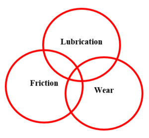
Overlapping key fields of study in Tribology
Tribology is mainly composed of three fields, namely friction, wear, and lubrication. These terms can be defined as follows:
Friction: Friction is the resistance to relative motion between two bodies in contact. There are two microscopic forces, called molecular adhesion and mechanical abrasion, between these two objects in touch. Molecular adhesion includes electrostatic, Van der Waals, as well as metallic bonds while mechanical abrasion comprises elastic, plastic, and viscoelastic deformations. Friction can be measured by the coefficient of friction which is a constant of proportionality.
Wear: Wear is the removal or displacement of material from one body when subjected to contact and relative motion with another body. Like friction, wear is a system property and not a material property. There are several distinct wear regimes, including abrasive wear, adhesive wear, fretting corrosion, erosive wear, rolling contact fatigue, and tribo-corrosion. Some of these regimes can operate simultaneously or sequentially. The wear rate significantly changes according to the wear mode which is a function of the Tribosystem.
Lubrication: Lubrication is the use of a fluid to minimize friction and wear. The critical roles of a lubricant are to reduce friction, prevent or minimize wear, transport debris away from the interface, and provide cooling. Different lubrication regimes, including boundary lubrication, mixed lubrication, and fluid film lubrication, are determined based on the fluid film thickness. These regimes are also described by the Stribeck curve.
The expression of “Biotribology” was first defined by Dowson in 1970, which considers all aspects of tribology associated with biological systems, particularly the synovial joints and joint replacements. Biotribology pertains to friction, wear, and lubrication at biological interfaces. Studying the biotribology of the natural synovial joints helps us to better understand the joints’ function as well as the development of related diseases, and to figure out what kind of medical interventions are required. This could contribute to enhancing the quality of life of patients suffering from diseases associated with synovial joints.
This article was written by Mahdieh Mosayebias part of an ongoing series of scientific communications written and curated by BioTrib’s Early Stage Researchers.
Mahdieh is researching the Design of Self Lubricating Prothesis at ETH Zurich, Switzerland.
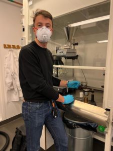
Since August, Vidhiaza and Giulio have been going to SWERIM AB as part of their BioTrib secondment plans. SWERIM AB is an institute that conducts industrial research and metals development. They have a location in Stockholm, just an hour away by train from Uppsala. At SWERIM AB, Vidhiaza and Giulio conduct experiments with the powder that they use for additive manufacturing. This brings a lot of synergy to their research as Uppsala University does not have the instruments to perform powder research.
Below, Vidhiaza is pictured with a spreadability tester in the SWERIM AB powder lab. This equipment can mimic the movement of a powder recoater that exists in an additive manufacturing machine to produce a layer of powder. Vidhiaza and Giulio use this machine to check and predict if their powder will be readily spreadable during the AM process, i.e., the powder produces a nice, dense layer.
Here Giulio is pictured with a setup for powder flowability measurements. SWERIM AB powder lab has different set ups for powder flowability according to different standards. Flowability is also an exciting and relevant powder property to AM process because it is directly related to how well an AM machine can dispense the powder.
Overall, the secondment at SWERIM AB has been very educative for Vidhiaza and Giulio, and it definitely enriches their research experience as BioTrib ESRs!
This article was written by Giulio Cavaliere as part of a series articles curated by BioTrib’s Early Stage Researchers.
Giulio Cavaliere is investigating Additively manufactured biodegradable alloys for bone replacement at Uppsala University, Sweden.
This article was written by Vidhiaza Leviandhika as part of an ongoing series of scientific communications written and curated by BioTrib’s Early Stage Researchers.
Vidhiaza is researching the Development of Development of 3D-printed gradient alloys for joint implant component at Uppsala University, Sweden
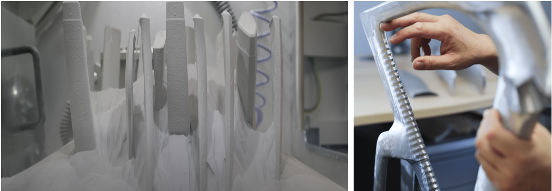
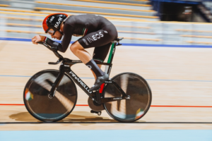
Header Image: 3D Printed Pinarello Bolide F HR 3D bike [1].
The ultimate benchmark for a cyclist, 60 minutes alone in a velodrome, suffering and draining any drop of energy left in the body. It’s the Hour record, the longest distance cycled in an hour from a stationary position.
From 1876, when the American Frank Dodds rode 26.508 km on a penny-farthing, the hour record has been held by the greatest cyclists in history from Fausto Coppi to Eddy Merckx, from Francesco Moser to Bradley Wiggins.
But the record is not just an incredible demonstration of human strength and endurance, it’s also the place to test and develop new cutting-edge technologies. In 1984 Francesco Moser smashed the record established 12 years before by Eddy Merckx using a revolutionary bike with composite lenticular wheels and a very peculiar frame shape.
Rider on Pinarello Bolide F HR 3D Bike [1].38 years later the hour record is once again Italian, Filippo Ganna cycled 56,792 Km in an hour in the Swiss velodrome of Grenchen. The bike he rode is almost as impressive as the record itself. The Pinarello Bolide F HR 3D was the first metal 3D-printed bike. The company used a high permeance aluminum alloy (scalalloy) 3D printed in an EOS M400, an industrial scale powder bed fusion (PBF) system. This was a proof of concept and used all the advantages of 3D printing over traditional manufacturing techniques. Firstly, the flexibility in design, the scale-like pattern on the seat tube greatly helps with the aerodynamic, but this is only obtainable through additive manufacturing. But also, fast prototyping allows customizing the frame to the need of any individual athlete without the need of realizing expensive and time-consuming mold for carbon fiber frames.
References
[1] “Bolide F HR 3D | En | Pinarello Global.” Accessed October 21, 2022. https://pinarello.com/global/en/bikes/road/competition/bolide-f-hr-3d/bolide-f-1.
This article was written by Giulio Cavaliere as part of a series articles curated by BioTrib’s Early Stage Researchers.
Giulio Cavaliere is investigating Additively manufactured biodegradable alloys for bone replacement at Uppsala University, Sweden.
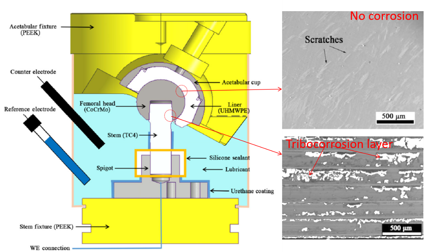
According to NJR (2021) report, UK continues to have the largest use of metal on polymer (MoP) hip replacements. An increasing number of the prevalence of revision surgery caused by adverse local tissue reaction in MoP THRs has recently been reported which is 3.2% at a mean follow-up of over 10 years (Hussey and McGrory, 2017). The issues are associated with debris/ion generated due to combined action of wear and corrosion termed as tribocorrosion/biotribocorrosion. As a result, the impact of these influential factors need to be assessed prior to implantation.
There are numbers of research questions related to hip implant failure which could be addressed during this preclinical testing using modern hip simulator. Such as, which interfaces are more prone to corrosion degradation? What is the tribocorrosion mechanism? etc. Often the results from preclinical testing showed deviance from clinical outcomes due to various reasons including testing protocol that is required to follow.
To address the above questions, Yang et al. (2022) have conducted an experiment under walking gait cycles to investigate the long-term biotribocorrosion at the MoP THR. His study revealed that the modular taper/trunnion junction was the main cause of MoP THR biotribocorrosion, not the bearing surfaces. The presence of tribochemical reaction layer on the surface proved the hypothesis as shown in the above figure (taken from the Yang et al. (2022) article and modified). The formation of this tribolayer could be impacted by number of ways. Even the changing of serum could have significant effect on the corrosion rate which may lead to poor prediction for clinical application. One of the limitation of his study is the lower number of loading cycles than recommended by ISO which need to be dealt in future.
Featured header image: Graphical abstract produced from figures taken from Yang et al. (2022)
References
Hussey D K, McGrory B J. Ten-year cross-sectional study of mechanically assisted crevice corrosion in 1,352 consecutive patients with metal-on-polyethylene total hip arthroplasty. J Arthroplasty 32(8): 2546–2551 (2017)
This article was written by MM Raihan as part of an ongoing series of scientific communications written and curated by BioTrib’s Early Stage Researchers.
Raihan is researching In-situ Measurement of Nano-scale Wear Utilising Advanced Sensors at the University of Leeds, UK.
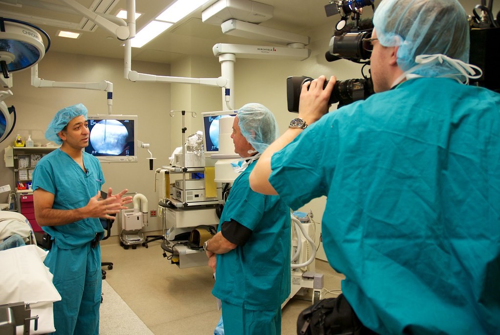
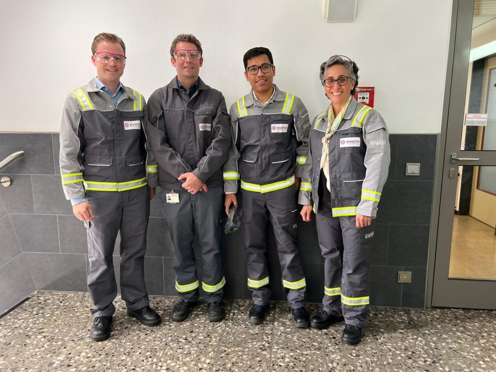
Picture: After visit to compounding floor, Marl (From left to right: Philip Engel (Head of Segment Medical Systems Europe, Evonik), Joscha Sandhusen (Compounding Technology, Evonik), Dilesh Raj Shrestha (PhD ESR, BioTrib-LTU), and Nazanin Emami (Professor, LTU)
I recently had the opportunity to visit two Evonik Industries AG sites in Germany (Marl and Darmstadt) as part of my industrial secondment. Evonik is one of the world’s largest specialty chemical companies, with a diverse business portfolio. Evonik is also active in the medical industry, manufacturing high-performance plastics like Poly Ether Ether Ketone (PEEK) for various implant applications. Evonik located at Marl Chemical Park, Evonik’s largest chemical park, where I spent my first week learning PEEK polymer processing and compounding. We were able to meet with experts in PEEK compounding development and have an in-depth discussion about current challenges and prospects.
I relocated to Evonik Darmstadt for the final three weeks of my secondment. In Darmstadt, Evonik has a tribology lab dedicated for material screening. I worked on the tribology test setup and wear data interpretation for medical implant applications. This provided me with an understanding of the clinically relevant tribological test setup required for material screening as well as the data interpretation techniques. I had the opportunity to meet with and speak with the medical implant team, where I learned about the industrial approach as well as their thoughts on moving forward with PEEK medical implant applications.
Finally, I’d like to express my gratitude to everyone who assisted in organizing this secondment and making it a fruitful learning experience.
This article was written by Dilesh Raj Shrestha as part of an ongoing series of scientific communications written and curated by BioTrib’s Early Stage Researchers.
Dilesh is researching the Development of 3D-printable, self-lubricated polymer composites with improved wear resistance for total joint replacement at Luleå University of Technology, Sweden.
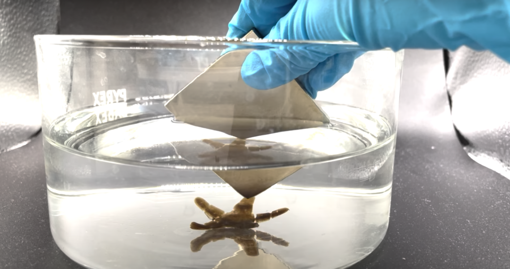
A study done by Julia et al. proved the possibility of using multi-material direct ink printing (4D printing) technology to integrate magnetic nanoparticles with hydrogels. The contactless in-air control of motions like rolling, jumping, and bending is realised because of the interactions between magnetic and nonmagnetic hydrogels. The programmability of patterns of the multi-material also enables its future potential in soft robotics
Starfish 3D printed responsive hydrogels by Dr Ali Mohammed:
Image of Starfish shaped responsive Hydrogel reporoduced with permission from Dr Ali Mohammed
Another fantastic work of 3D printed responsive hydrogels by Dr Ali Mohammed!
The superparamagnetic starfish-shaped hydrogel can be used for magnetically simulated soft robotics and actuators.
Check out the videos to see the 3D printed ‘starfish’ moving responsively to the magnet:
This post was written by Esperanza Shi as part of an ongoing series of scientific communications written and curated by BioTrib’s Early Stage Researchers.
Esperanza is researching the Optimisation of Scanning Strategies for 3D Printed Artificial Joints at Imperial College London, UK.
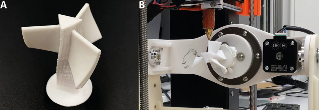
Turbine-like object printed by 5-axis 3d printing [1]
To improve the quality of printed overhanging structures and surfaces, to reduce the waste of materials of support structures and to reduce the printing time, 3D printers are ready to evolve into multi-axis. Freddie Hong, a PhD candidate at Imperial College London, has been designing 5-axis 3D printers and conformal slicing in an accessible and cheap way to bring these advantages to more individuals. With the rotating printing platform and the slicer designed for 5-axis 3D printers, the overhanging structures can be printed conformally, resulting in reinforced structural strengths.
Visit the video below to watch Freddie sharing more details!
Freddie made the hardware and software kits of open5x open resource, so check out the GitHub and upgrade your desktop 3D printer!
[1] Hong, Freddie, et al. “Open5x: Accessible 5-axis 3D printing and conformal slicing.” CHI Conference on Human Factors in Computing Systems Extended Abstracts. 2022.
This post was written by Esperanza Shi as part of an ongoing series of scientific communications written and curated by BioTrib’s Early Stage Researchers.
Esperanza is researching the Optimisation of Scanning Strategies for 3D Printed Artificial Joints at Imperial College London, UK.
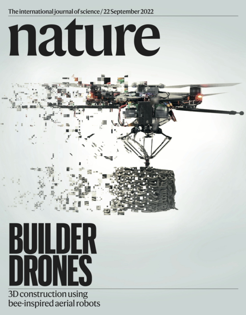
Breaking news from Imperial College London and Empa Switzerland that the research of flying 3D printers is featured as the cover of Nature (Volume 609 Issue 7928, 22 September 2022). Instead of sitting statically on the table like most 3d printers, this aerial robotic 3d printer prints structures in-flight, inspired by natural builders like wasps and bees.
The ‘ScanDrones’ work in pairs with the ‘BuildDrones’ enabling the monitoring of the print quality, thanks to a generic real-time model-predictive-control scheme. Flying 3D printers have been proven to have the potential in conducting constructions post-disaster or in places that are difficult to access. Check out the video produced by the Imperial and Empa researchers below:
Visit the website and original paper below to find out more!
Zhang, Ketao, et al. “Aerial additive manufacturing with multiple autonomous robots.” Nature 609.7928 (2022): 709-717.
This post was written by Esperanza Shi as part of an ongoing series of scientific communications written and curated by BioTrib’s Early Stage Researchers.
Esperanza is researching the Optimisation of Scanning Strategies for 3D Printed Artificial Joints at Imperial College London, UK.
Having been asked this question several times when I was trying to introduce myself and what I do with my PhD to someone I just met, I feel it’s time to update the general public on how cutting-edge research about this technology is looking like now. The fact is that the public no longer views 3D printing as ‘cutting-edge’ because 3D printing technology came out decades ago and has become so easy to access (you can easily buy one desktop 3D printer and set it up at home).
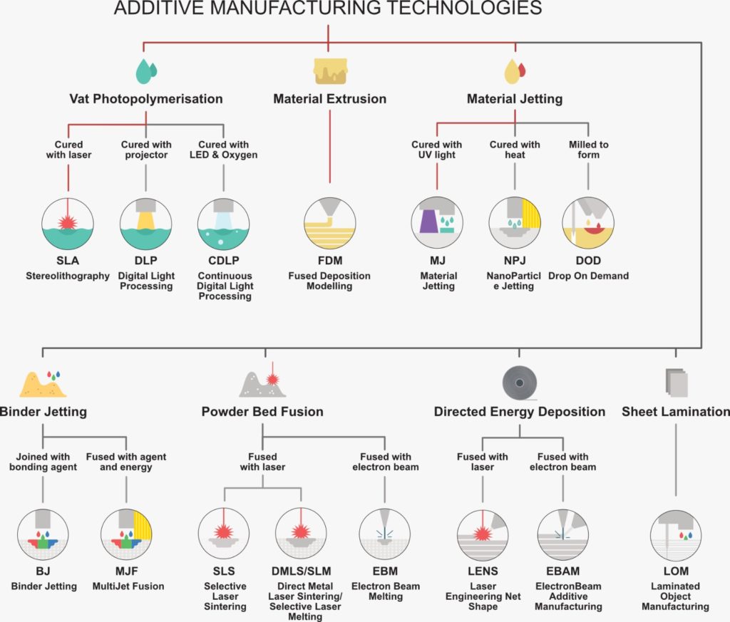
However, 3D printing is not just about the desktop 3D printer you can have at home that extrudes plastics. Just have a glance at this map, and you will understand that 3D printing is such a broad concept and there is much more it’s capable of.
Picture by Dr Usman Waheed
There’re so many aspects we can improve under each category to make the technology better, meanwhile, engineers and scientists never stopped exploring the potential and broadening the horizon of 3D printing. The map or we can say the world of 3D printing is still getting bigger and better, so we DO need PhDs in 3D printing!
I’m not saying this because I’m trying to defend myself as personally being a PhD in 3D printing. The 3 amazing examples I’m going to show you over the next two weeks are to convince you that we DO need PhDs in 3D printing.
This post was written by Esperanza Shi as part of an ongoing series of scientific communications written and curated by BioTrib’s Early Stage Researchers.
Esperanza is researching the Optimisation of Scanning Strategies for 3D Printed Artificial Joints at Imperial College London, UK.
About a year ago, to pursue my academic career I moved from Italy to Sweden, specifically to Uppsala, a lovely town near Stockholm. I am now a PhD student in the local university and in some way I feel that my undergraduate days are well behind me. But you know, Uppsala is a very special city full of students from all around the world who certainly “rule” the town. Here, even if you are no longer a student, you can easily feel like one again just walking into a specific neighborhood at a specific time of the day… Or should I say night?
This neighborhood is called Flogsta, it is located in the west part of the city and most of its inhabitants (basically everyone) are students at Uppsala University. But what’s special about this place you might ask? There, every evening at about 10 p.m. the “Flogsta Scream” can be heard. If you’re from Uppsala you will surely be familiar with this particular “ritual”, or at least have heard of it. Literally. If instead you don’t know anything about it, the Flogsta Scream is (as the name suggest) a scream, but it’s also something more than that. It is a collective act in which students scream together from windows, balconies and rooftops. According to the student population, this act is a kind of “safety valve” or “a cry of anguish” over the accumulated stress of the demands of college life.
Truth be told, it is just an occasion to socialize and make some noise together, but for Uppsala students it is something that you can count on every single day. Sometimes everyone need to scream a little in life, might as well do it the Uppsala way.
This article was written by Niccoló De Berardinis as part of an ongoing series of scientific communications written and curated by BioTrib’s Early Stage Researchers.
Niccoló is researching Bioimaging of biomaterials and biological characterization of 3D-printed alloys for reconstructive surgery at Uppsala University, Sweden.
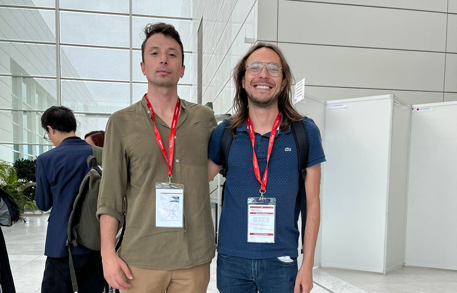
From the 4th to the 8th of September the European Society of Biomaterials (ESB) hosted its 32nd conference at Palais des Congrès, in Bordeaux, France. ESRs André Plath (ETH Zürich) and Giulio Cavaliere (U. Uppsala) presented some of their current results in poster format during the conference.
“The conference was a great opportunity for networking and building a strong basis for future collaborations. The plenary talks were not only inspiring but represented the top-notch science being developed right now. It was also great to see a good mixture of academics and industries. I commend the ESB for also organizing lunches and talks with senior researchers, industrials, and editors.” says André.
”We met plenty of people working on similar projects and it was a great context to exchange ideas and get insightful feedbacks on my work. Being my first conference I also learnt a lot of useful skills on how to present my research. A lot of the plenary talks were more focus on tissues engineering so it was a good opportunity to expand my knowledge outside my research field” says Giulio
This article was written by André Plath and Giulio Cavaliere as part of a series articles curated by BioTrib’s Early Stage Researchers.
André is one of BioTrib’s Early Stage Researcher‘s who is investigating Boundary Lubrication of Fibrous Scaffolds at ETH Zürich, Switzerland.
Giulio Cavaliere is investigating Additively manufactured biodegradable alloys for bone replacement at Uppsala University, Sweden.